I. TISSUES — OVERVIEW
A TWO MAIN GROUPS (BASED ON DIVIDING POWER)
| TYPE | CAPACITY TO DIVIDE | DETAIL |
|---|---|---|
| Meristematic tissue | Can divide (retain capacity throughout life) | Growth regions |
| Permanent tissue | Cannot divide (lost dividing capacity) | Differentiated, mature |
NEET 2021: Tissues are classified into meristematic & permanent based on their division power.
II. MERISTEMATIC TISSUES
A CLASSIFICATION BASED ON POSITION
| TYPE | LOCATION | TISSUE PRODUCED | DETAIL |
|---|---|---|---|
| Apical meristems | Tips of roots & shoots | Primary tissues | Responsible for primary growth (increase in length) |
| Intercalary meristems | Between mature tissues (e.g., base of internodes) | Primary tissues | Occur in grasses; regenerate parts removed by grazing herbivores ⭐ |
| Lateral meristems | Along the sides (cylindrical) | Secondary tissues | Responsible for secondary growth (increase in girth) |
NEET 2010: Both apical & intercalary meristems are PRIMARY meristems.
NEET 2023: Axillary buds and intercalary meristems = primary meristems.
Lateral Meristems — Examples ⭐
| EXAMPLE |
|---|
| Fascicular vascular cambium |
| Interfascicular cambium |
| Cork cambium (Phellogen) |
B VASCULAR CAMBIUM — ORIGIN
| PLANT PART | ORIGIN OF VASCULAR CAMBIUM |
|---|---|
| Dicot stem | Partly primary (fascicular) & partly secondary (interfascicular) |
| Dicot root | Completely secondary in origin ⭐ |
III. PERMANENT TISSUES
| TYPE | DESCRIPTION |
|---|---|
| Simple tissue | All cells similar in structure & function |
| Complex tissue | Many different types of cells working together |
NEET 2021: Permanent tissues — cells do not divide further; simple (similar cells) vs complex (different cells).
A SIMPLE TISSUES — MASTER COMPARISON TABLE
| FEATURE | PARENCHYMA | COLLENCHYMA | SCLERENCHYMA |
|---|---|---|---|
| Cell shape | Generally isodiametric; various shapes | — | Long, narrow |
| Cell wall | Thin, cellulosic | Thickened at corners (unevenly) due to cellulose, hemicellulose & pectin ⭐ | Thick, lignified walls with pits |
| Living / Dead | Living | Living ⭐⭐ | Dead (without protoplast) ⭐ |
| Intercellular spaces | Closely packed OR with small spaces | Absent ⭐ | — |
| Mechanical support | No (turgidity provides some) | Yes — Living mechanical tissue ⭐ | Yes — Non-living mechanical tissue ⭐ |
| Location | Cortex, pericycle, pith, medullary rays | Hypodermis of dicot plants ⭐ | Groups; throughout plant body |
| Functions | Photosynthesis, food storage, secretion | Mechanical strength to young stems | Mechanical support |
NEET 2021, 2024: Parenchyma = simple, living tissue; all cells similar.
NEET 2024 TRAP: Collenchyma is LIVING tissue (statement saying 'collenchyma is dead' is FALSE!).
SCLERENCHYMA — Two Types ⭐⭐
| TYPE | SHAPE | FEATURES | FOUND IN |
|---|---|---|---|
| Fibres | Thick-walled, elongated, pointed; occur in groups | Long cells | Various plant parts |
| Sclereids | Spherical, oval or cylindrical | Highly thickened dead cells; very narrow lumen ⭐ | Fruit walls of nuts; pulp of guava / pear / sapota; seed coat of legumes; leaves of tea ⭐ |
NEET 2021, 2022: Sclereids = dead, highly thickened, narrow lumen.
RE-NEET 2024: Sclereids found in fruit walls of nuts, pulp of guava / pear / sapota, seed coat of legumes, leaves of tea.
B COMPLEX TISSUES — XYLEM (Water & Mineral Conducting)
| FEATURE | DETAIL |
|---|---|
| Function | Conducts water & minerals from roots → stem → leaves |
| Four elements | Tracheids, Vessels, Xylem fibres, Xylem parenchyma |
Xylem Elements — Detailed ⭐⭐
| ELEMENT | LIVING / DEAD | KEY FEATURES |
|---|---|---|
| Tracheids | Dead | Unicellular; elongated; imperforate (no perforation plates) ⭐; present in ALL vascular plants |
| Vessels | Dead | Multicellular (long cylindrical tube-like); characteristic of Angiosperms ⭐; absent in Gymnosperms ⭐⭐ |
| Xylem fibres | Dead | — |
| Xylem parenchyma | Living ⭐ | Stores food as starch, fats & tannins; radial conduction of water by ray parenchyma |
NEET 2010, 2014: Tracheids & vessels = main water-transporting elements in flowering plants. Tracheids are unicellular; vessels are multicellular. Tracheids differ from vessels by being imperforate (no perforation plates).
NEET 2012, 2019, RE-NEET 2024: Vessels are characteristic of Angiosperms and absent in Gymnosperms!
Primary Xylem — Two Types ⭐⭐
| TYPE | DETAIL |
|---|---|
| Protoxylem | First formed xylem |
| Metaxylem | Later formed xylem |
Endarch vs Exarch ⭐⭐⭐
| ARRANGEMENT | PROTOXYLEM POSITION | FOUND IN |
|---|---|---|
| Endarch | Protoxylem towards the centre (pith) | Stems ⭐ |
| Exarch | Protoxylem towards the periphery | Roots ⭐ |
NEET 2023: Endarch protoxylem in stems; Exarch protoxylem in roots.
NEET 2014: To distinguish old stem from root → look at position of protoxylem (endarch = stem; exarch = root).
Tyloses ⭐
NEET 2016: Tyloses = extensions of xylem parenchyma into vessels in heartwood.
C COMPLEX TISSUES — PHLOEM (Food Conducting)
| FEATURE | DETAIL |
|---|---|
| Function | Transports food materials (photosynthates) usually from leaves → other parts |
| Four elements | Sieve tube elements, Companion cells, Phloem parenchyma, Phloem fibres |
Phloem Elements — Detailed ⭐⭐
| ELEMENT | LIVING / DEAD | KEY FEATURES |
|---|---|---|
| Sieve tube elements | Living | Mature sieve tubes lack nucleus ⭐; function controlled by nucleus of companion cells |
| Companion cells | Living | Specialised parenchymatous cells; closely associated with sieve tube elements ⭐; help maintain pressure gradient in sieve tubes |
| Phloem parenchyma | Living | Stores food, resin, latex, mucilage; absent in most monocotyledons ⭐⭐ |
| Phloem fibres (Bast fibres) | Dead ⭐ | Made of sclerenchymatous cells; absent in primary phloem but present in secondary phloem |
NEET 2012: Companion cells = specialised parenchymatous cells closely associated with sieve tubes.
NEET 2024: Companion cells help in maintenance of pressure gradient in sieve tubes.
Gymnosperms vs Angiosperms — Phloem ⭐⭐
| FEATURE | GYMNOSPERMS | ANGIOSPERMS |
|---|---|---|
| Sieve tube elements | Absent | Present |
| Companion cells | Absent | Present |
| Instead they have | Sieve cells + Albuminous cells | — |
NEET 2012, 2019, RE-NEET 2024: Gymnosperms lack sieve tubes & companion cells → have albuminous cells & sieve cells instead.
GYMNOSPERM vs ANGIOSPERM — QUICK COMPARISON (Xylem + Phloem)
| FEATURE | GYMNOSPERMS | ANGIOSPERMS |
|---|---|---|
| Xylem vessels | Absent ⭐ | Present |
| Sieve tube elements | Absent | Present |
| Companion cells | Absent | Present |
| Present instead | Tracheids (xylem); Sieve cells + Albuminous cells (phloem) | All elements present |
IV. THE TISSUE SYSTEM
Three types of tissue systems based on structure & location:
| SYSTEM | COMPONENTS |
|---|---|
| 1. Epidermal tissue system | Epidermal cells, Stomata, Epidermal appendages (trichomes & hairs) |
| 2. Ground / Fundamental tissue system | Parenchyma, Collenchyma, Sclerenchyma (cortex, pericycle, pith, mesophyll) |
| 3. Vascular tissue system | Xylem + Phloem (vascular bundles) |
A EPIDERMAL TISSUE SYSTEM
General Features ⭐
| FEATURE | DETAIL |
|---|---|
| Position | Outermost covering of whole plant body |
| Layers | Usually single-layered |
| Cell type | Parenchymatous; elongated, compactly arranged, continuous layer |
| Cytoplasm | Small amount; cell wall is lined by it |
| Vacuole | Large |
| Cuticle | Waxy thick layer on outside → prevents water loss ⭐ |
| Cuticle in roots | Absent ⭐ |
Stomata ⭐⭐⭐
| FEATURE | DETAIL |
|---|---|
| Function | Regulate transpiration & gaseous exchange |
| Guard cells (Dicots) | Two bean-shaped cells ⭐ |
| Guard cells (Monocots) | Dumb-bell shaped (grasses) ⭐ |
| Guard cell walls | Outer walls (away from pore) = thin; Inner walls (towards pore) = highly thickened |
| Chloroplasts | Guard cells possess chloroplasts ⭐ |
| Subsidiary cells | Specialised epidermal cells surrounding guard cells (in shape & size) |
NEET 2011, 2016, 2018, 2024, 2026 (reinforced): Guard cells contain chloroplasts in land plants; Dumb-bell shaped guard cells in monocots (grasses); Subsidiary cells = specialised cells in vicinity of guard cells.
Stomata Structure & Guard Cells
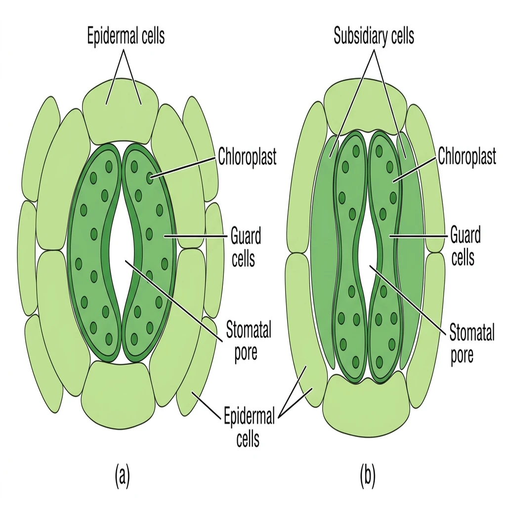
Structure of Stomata: Shows epidermal pores regulated by bean-shaped guard cells (dicots) and dumb-bell shaped guard cells (monocots).
🔬 Detailed Biochemical & Structural Description
Stomata are microscopic structures present in the epidermis of leaves regulating transpiration and gas exchange. Each stoma is composed of a stomatal pore guarded by two specialized 'Guard cells' containing 'Chloroplasts'. (a) Dicotyledons possess bean-shaped guard cells flanked by subsidiary cells. (b) Monocotyledons (grasses) possess dumb-bell shaped guard cells with vertical walls.
Epidermal Hairs ⭐
| TYPE | FEATURES | FUNCTION |
|---|---|---|
| Root hairs | Unicellular elongations of epidermal cells | Absorb water & minerals from soil |
| Trichomes (Stem hairs) | Usually multicellular; branched or unbranched; soft or stiff; may be secretory | Prevent water loss due to transpiration ⭐ |
NEET 2011, 2026 (reinforced): Root hairs = unicellular; arise from region of maturation (tubular extensions of epidermal cells). Trichomes = usually multicellular.
B GROUND TISSUE SYSTEM
• All tissues except epidermis and vascular bundles = ground tissue
• Consists of parenchyma, collenchyma & sclerenchyma
• Present in cortex, pericycle, pith, medullary rays
• ⭐ In leaves, ground tissue = mesophyll (chloroplast-containing parenchyma)
NEET 2021: Mesophyll = ground tissue of leaves = chloroplast-containing.
C VASCULAR TISSUE SYSTEM
Types of Vascular Bundles ⭐⭐
| FEATURE | OPEN | CLOSED |
|---|---|---|
| Cambium | Present (between xylem & phloem) | Absent |
| Secondary growth | Can form secondary xylem & phloem | Cannot form secondary tissues |
| Found in | Dicot stems ⭐ | Monocot stems ⭐ |
| FEATURE | RADIAL | CONJOINT |
|---|---|---|
| Arrangement | Xylem & phloem at alternate radii | Xylem & phloem on same radius |
| Found in | Roots ⭐ | Stems & Leaves ⭐ |
| Phloem position | — | Usually on outer side of xylem |
NEET 2012, 2018, 2019: Open vascular bundles in dicot stem (cambium present); Closed in monocot stem (cambium absent).
NEET 2022: Radial vascular bundles in roots; Conjoint in stems / leaves.
Types of Vascular Bundles
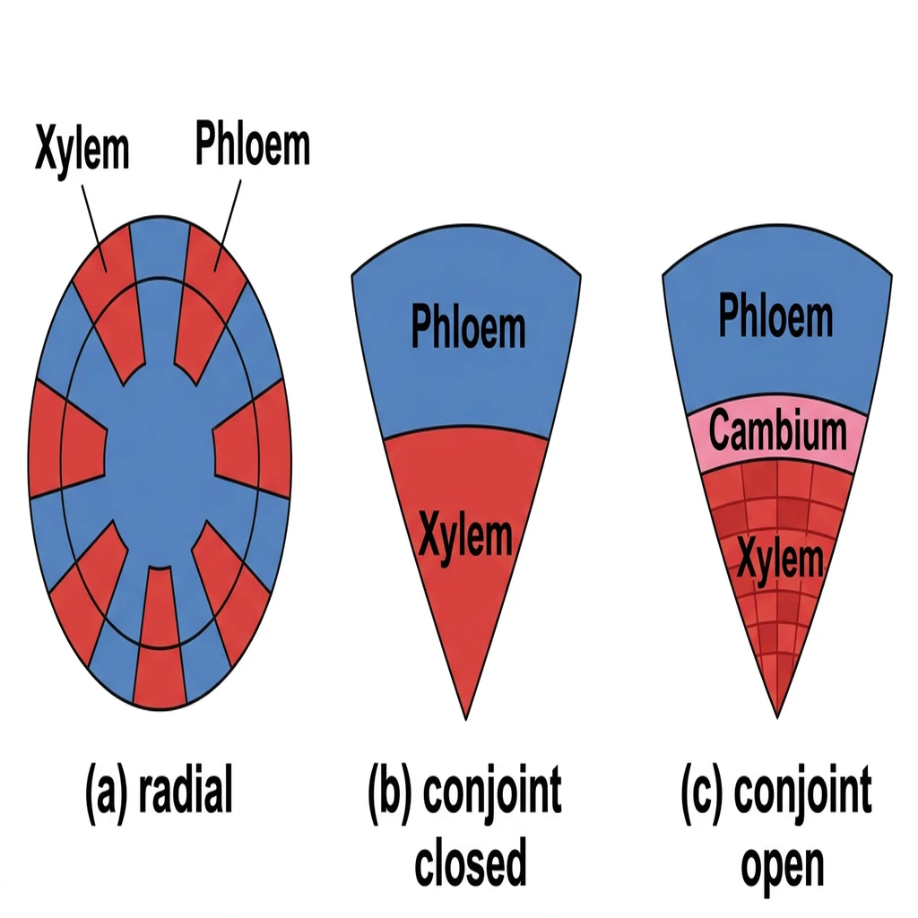
Vascular Bundle Formations: Compares radial (roots) and conjoint (stems/leaves) closed and open tissue configurations.
🔬 Detailed Biochemical & Structural Description
Vascular bundles consist of xylem and phloem complex tissues, classified into: (a) Radial: Xylem and phloem occur in alternating patches on different radii (typical of roots); (b) Conjoint Closed: Xylem and phloem occur on the same radius without intermediate cambium (typical of monocot stems); (c) Conjoint Open: Strip of meristematic 'Cambium' is present between xylem and phloem, enabling secondary growth (typical of dicot stems).
V. ANATOMY OF ROOTS, STEMS & LEAVES
A DICOT ROOT ⭐⭐
| LAYER (OUTSIDE → INSIDE) | FEATURES |
|---|---|
| Epiblema (Epidermis) | Outermost layer; many cells protrude as unicellular root hairs |
| Cortex | Several layers of thin-walled parenchyma cells with intercellular spaces |
| Endodermis ⭐⭐ (innermost cortex) | Single layer of barrel-shaped cells; no intercellular spaces; tangential & radial walls have Casparian strips (waxy, water-impermeable material = suberin) ⭐⭐ |
| Pericycle | Few layers of thick-walled parenchymatous cells; initiates lateral roots & vascular cambium during secondary growth ⭐ |
| Vascular bundles | 2–4 xylem & phloem patches; arranged in radial pattern; exarch protoxylem ⭐ |
| Conjunctive tissue | Parenchymatous cells between xylem & phloem |
| Pith | Small or inconspicuous ⭐ |
NEET 2018, 2026 (reinforced): Casparian strips = deposition of suberin (waxy, water-impermeable) on radial & tangential walls of endodermal cells in roots. Conjunctive tissue is tissue between xylem and phloem!
NEET 2016: Cortex = region between epidermis and stele (endodermis is innermost layer of cortex).
B MONOCOT ROOT ⭐
| FEATURE | DETAIL |
|---|---|
| Overall | Similar to dicot root in many respects |
| Epiblema, cortex, endodermis, pericycle | Same as dicot root |
| Xylem bundles | More than six = Polyarch condition ⭐⭐ |
| Pith | Large & well developed ⭐ (contrast: dicot root pith is small) |
| Secondary growth | Absent ⭐ |
| Vascular arrangement | Radial; exarch protoxylem |
NEET 2015, 2022, 2023: Monocot root = polyarch, radial, exarch, large pith, no cambium, no secondary growth.
Anatomy of Dicot and Monocot Roots
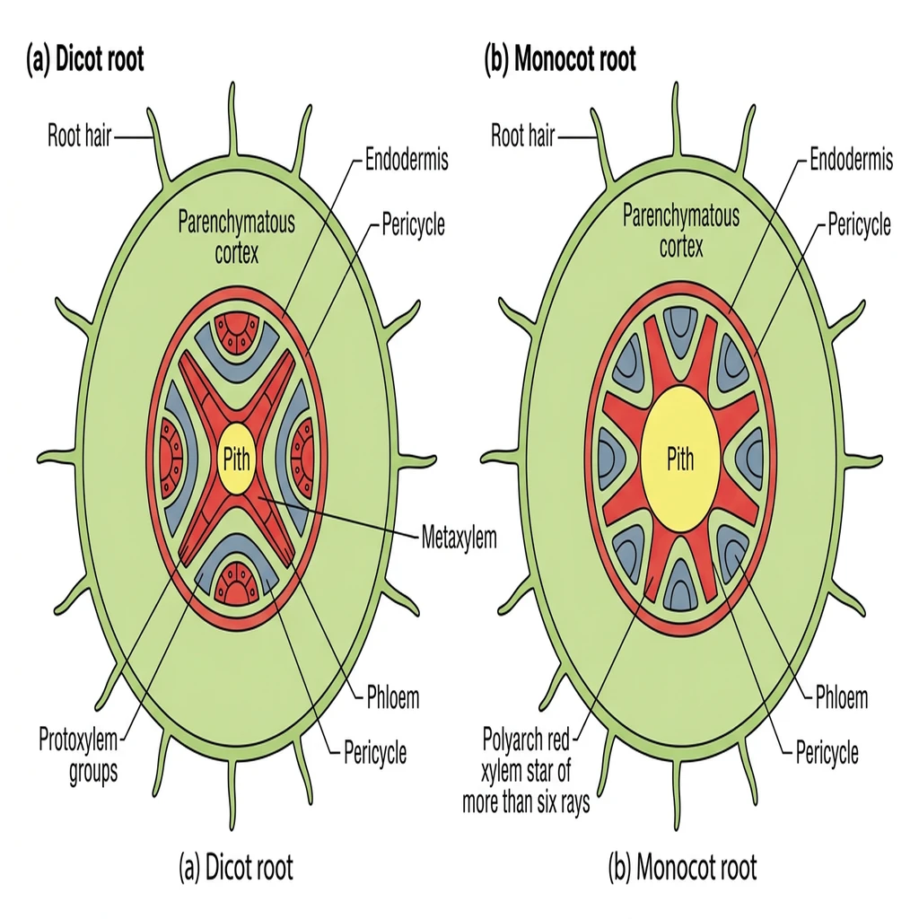
Root T.S. Comparison: Highlights cortex, endodermis, pericycle, and xylem/phloem arrangements in dicot vs. monocot roots.
🔬 Detailed Biochemical & Structural Description
Transverse sections (T.S.) of roots highlight tissue systems: (a) Dicot Root: Features unicellular root hairs, cortex parenchyma, a distinct endodermis with suberized Casparian strips, pericycle, and diarch to tetrarch xylem bundles with small central pith; (b) Monocot Root: Similar ground tissues but features a polyarch xylem (six or more bundles) and a large, well-developed central pith.
C DICOT STEM ⭐⭐⭐
| LAYER (OUTSIDE → INSIDE) | FEATURES |
|---|---|
| Epidermis | Outermost protective layer; covered with thin cuticle; may bear trichomes & few stomata |
| Cortex (3 sub-zones) | See below |
| Endodermis | Innermost cortex layer; cells rich in starch grains → called 'starch sheath' ⭐ |
| Pericycle | On inner side of endodermis; semi-lunar patches of sclerenchyma above the phloem ⭐ |
| Medullary rays | Radially placed parenchymatous cells between vascular bundles |
| Vascular bundles | Arranged in a ring ⭐⭐ (characteristic of dicot stem); conjoint, open, endarch protoxylem ⭐ |
| Pith | Central portion; large, rounded parenchymatous cells with large intercellular spaces |
Cortex Sub-zones ⭐
| SUB-ZONE | TISSUE | DETAIL |
|---|---|---|
| Hypodermis (outer) | Collenchymatous ⭐⭐ | Few layers just below epidermis; provides mechanical strength |
| Cortical layers (middle) | Parenchymatous | Rounded thin-walled cells with conspicuous intercellular spaces |
| Endodermis (inner) | — | Starch sheath |
NEET 2020, 2026 (reinforced): Dicot stem — vascular bundles arranged in ring; conjoint, open, endarch. Starch sheath is endodermal cells rich in starch grains — innermost layer of cortex in dicot stem!
D MONOCOT STEM ⭐⭐⭐
| FEATURE | DETAIL |
|---|---|
| Hypodermis | Sclerenchymatous ⭐⭐ (contrast: dicot = collenchymatous) |
| Vascular bundles | Scattered (not in a ring) ⭐⭐; each surrounded by sclerenchymatous bundle sheath |
| VB type | Conjoint & closed ⭐ (no cambium → no secondary growth) |
| Bundle size | Peripheral bundles = smaller; Central bundles = larger |
| Phloem parenchyma | Absent ⭐⭐ |
| Water-containing cavities | Present within vascular bundles ⭐ |
| Ground tissue | Large, conspicuous, parenchymatous |
| Endarch protoxylem | Yes |
NEET 2012, 2020, 2025 (reinforced): Monocot stem — hypodermis is sclerenchymatous (not parenchymatous!); scattered vascular bundles, sclerenchymatous bundle sheath, conjoint & closed; phloem parenchyma absent; water-containing cavities present.
E DICOT STEM vs MONOCOT STEM — COMPARISON
| FEATURE | DICOT STEM | MONOCOT STEM |
|---|---|---|
| Hypodermis | Collenchymatous | Sclerenchymatous |
| VB arrangement | Arranged in a ring | Scattered |
| Bundle sheath | — | Sclerenchymatous |
| VB type | Conjoint, Open | Conjoint, Closed |
| Cambium | Present | Absent |
| Secondary growth | Occurs | Does not occur |
| Phloem parenchyma | Present | Absent |
| Medullary rays | Present | Absent |
| Pith | Present, distinct | Not distinguishable (ground tissue) |
| Endodermis | Starch sheath | Not distinct |
| Pericycle | Semi-lunar sclerenchyma patches | Not distinct |
F DICOT ROOT vs DICOT STEM — COMPARISON
| FEATURE | DICOT ROOT | DICOT STEM |
|---|---|---|
| Protoxylem position | Exarch (towards periphery) ⭐ | Endarch (towards centre / pith) ⭐ |
| VB type | Radial | Conjoint, Open |
| Endodermis | Casparian strips (suberin) | Starch sheath (starch grains) |
| Pericycle | Parenchymatous; initiates lateral roots | Sclerenchymatous (semi-lunar patches) |
| Pith | Small / inconspicuous | Large |
| Trichomes / hairs | Root hairs (unicellular) | Trichomes (multicellular) |
| Cuticle | Absent | Present |
Anatomy of Dicot and Monocot Stems
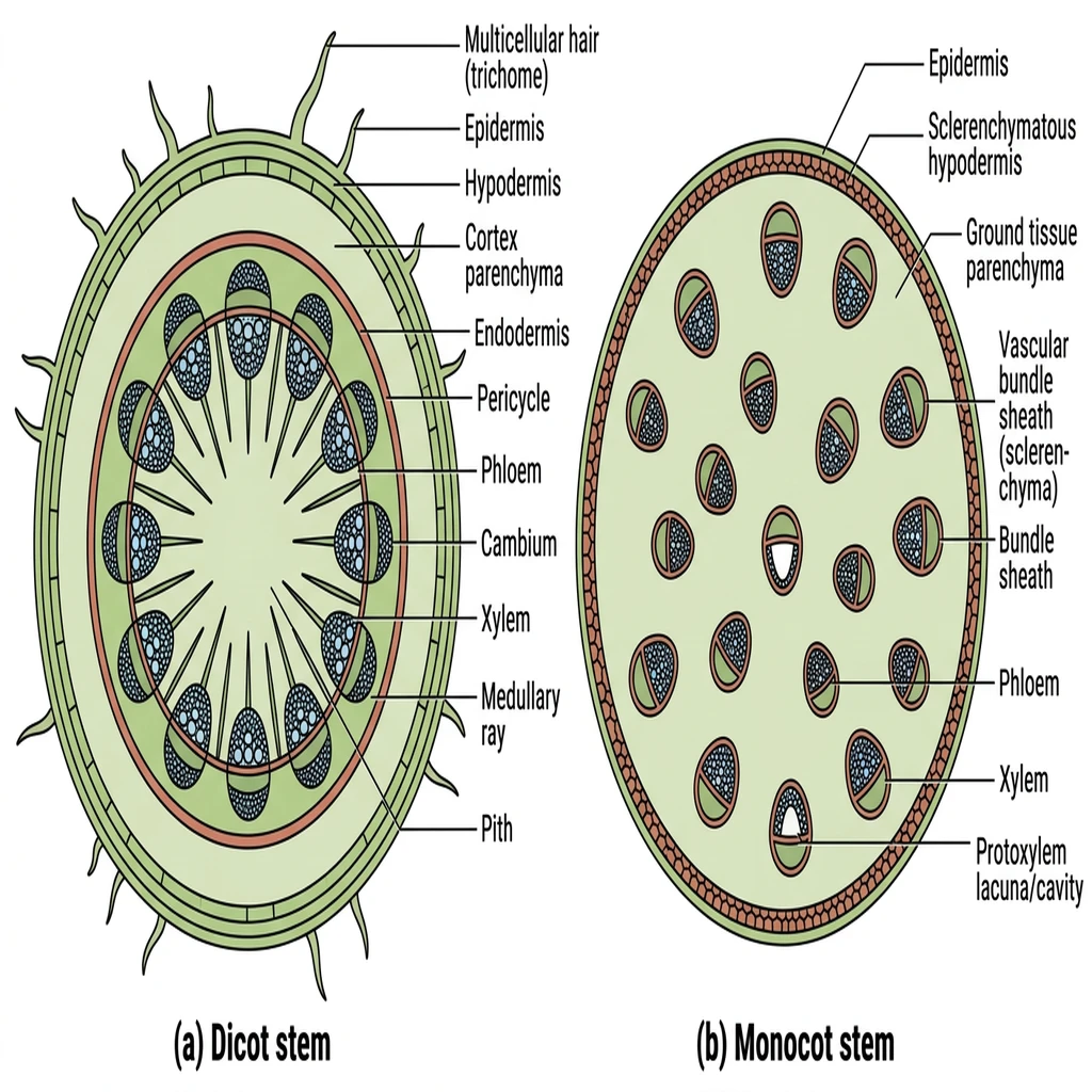
Stem T.S. Comparison: Contrasts the ringed, open vascular bundles of dicot stems with the scattered, closed bundles of monocot stems.
🔬 Detailed Biochemical & Structural Description
Stem anatomy contrasts tissue architectures: (a) Dicot Stem: Comprises epidermis with multicellular trichomes, collenchyma hypodermis, ring-arranged conjoint open vascular bundles capped by sclerenchyma pericycle, medullary rays, and a central parenchymatous pith; (b) Monocot Stem: Features a sclerenchymatous hypodermis, a massive undifferentiated ground tissue, and numerous scattered conjoint closed vascular bundles surrounded by sclerenchymatous bundle sheaths.
G DICOT LEAF (Dorsiventral Leaf) ⭐⭐
| FEATURE | DETAIL |
|---|---|
| Leaf type | Dorsiventral (different upper & lower surfaces) |
| Epidermis | Covers both surfaces; conspicuous cuticle |
| Stomata distribution | More on abaxial (lower) surface ⭐; adaxial may even lack stomata |
| Guard cells | Bean-shaped (dicot) ⭐ |
Mesophyll (Differentiated into Two Types) ⭐⭐
| TYPE | POSITION | CELL SHAPE | ARRANGEMENT |
|---|---|---|---|
| Palisade parenchyma | Adaxial (upper) surface ⭐ | Elongated cells | Vertically arranged, parallel to each other |
| Spongy parenchyma | Abaxial (lower) surface | Oval or round cells | Loosely arranged; numerous air cavities between cells |
• Vascular System: Vascular bundles in veins & midrib; size depends on size of veins (vary in thickness — reticulate venation); surrounded by layer of thick-walled bundle sheath cells.
RE-NEET 2024: Dicot leaf — dorsiventral; stomata more on abaxial surface; palisade parenchyma on adaxial surface, elongated, vertically arranged.
NEET 2024 TRAP: Statement saying 'adaxial epidermis bears more stomata' is FALSE — it's the abaxial that bears more!
H MONOCOT LEAF (Isobilateral Leaf) ⭐⭐
| FEATURE | DETAIL |
|---|---|
| Leaf type | Isobilateral (both surfaces similar) |
| Stomata | Present on both surfaces of epidermis (equal distribution) ⭐ |
| Guard cells | Dumb-bell shaped ⭐ |
| Mesophyll | NOT differentiated into palisade & spongy ⭐⭐ |
| Venation | Parallel → vascular bundles of near similar sizes (except main veins) |
Bulliform Cells ⭐⭐⭐
| FEATURE | DETAIL |
|---|---|
| Location | Adaxial epidermis (along the veins) in grasses |
| Cell type | Large, empty, colourless (modified adaxial epidermal cells) ⭐ |
| When turgid (absorbed water) | Leaf surface is exposed (leaf opens / flattens) |
| When flaccid (water stress) | Leaves curl inwards → minimise water loss ⭐⭐ |
NEET 2018, 2019, 2024, 2026 (reinforced): Bulliform cells — present in grasses; large, empty, colourless; on adaxial surface; when flaccid during water stress → leaves curl inwards to minimise water loss!
I DICOT LEAF vs MONOCOT LEAF — COMPARISON
| FEATURE | DICOT LEAF (DORSIVENTRAL) | MONOCOT LEAF (ISOBILATERAL) |
|---|---|---|
| Stomata | More on abaxial (lower) surface | Equal on both surfaces |
| Guard cell shape | Bean-shaped | Dumb-bell shaped |
| Mesophyll | Differentiated (palisade + spongy) | NOT differentiated |
| Bulliform cells | Absent | Present (adaxial epidermis, grasses) |
| Venation | Reticulate (varied VB size) | Parallel (similar VB size) |
Anatomy of Dicot and Monocot Leaves
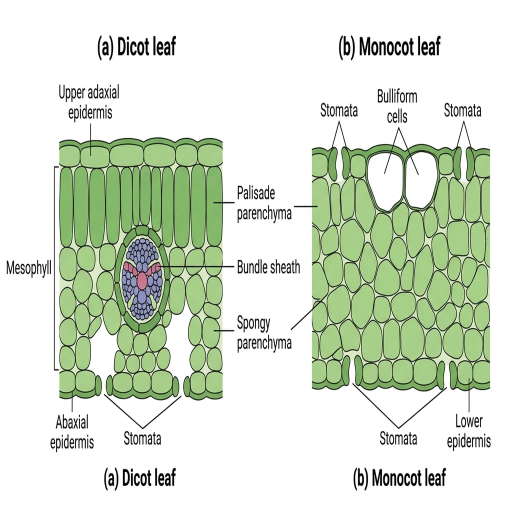
Leaf T.S. Comparison: Displays mesophyll differentiation (palisade vs. spongy) in dorsiventral dicot leaves vs. isobilateral monocot leaves.
🔬 Detailed Biochemical & Structural Description
Leaf internal structures differ by class: (a) Dorsiventral (Dicot) Leaf: Features distinct upper adaxial and lower abaxial epidermis, stomata mostly on the lower side, and mesophyll differentiated into upper palisade parenchyma and lower spongy parenchyma with air cavities; (b) Isobilateral (Monocot) Leaf: Features stomata on both sides, undifferentiated mesophyll, and large empty bulliform cells in the epidermis to regulate leaf rolling.
VI. SECONDARY GROWTH
Occurs in most dicotyledonous roots & stems. Absent in monocotyledons (e.g., grasses).
A VASCULAR CAMBIUM
• Formation in Dicot Stem: Interfascicular cambium develops from cells of medullary rays → joins the fascicular cambium → forms a complete cambium ring.
NEET 2013, 2022: Interfascicular cambium develops from medullary ray cells.
Activity of Vascular Cambium ⭐⭐
| DIRECTION | TISSUE PRODUCED |
|---|---|
| Inner side (towards pith) | Secondary xylem ⭐ |
| Outer side (towards cortex) | Secondary phloem ⭐ |
*More secondary xylem is produced than secondary phloem.*
Sequence from Pith → Cortex ⭐
NEET 2017, 2018: Vascular cambium cuts off secondary xylem on inner side and secondary phloem on outer side.
NEET 2023: Know the sequence: primary xylem → secondary xylem → cambium → secondary phloem → primary phloem.
B SPRING WOOD vs AUTUMN WOOD
| FEATURE | SPRING WOOD (EARLY WOOD) | AUTUMN WOOD (LATE WOOD) |
|---|---|---|
| Cambium activity | Very active | Less active |
| No. of xylem elements | Large number | Fewer |
| Vessel size | Wider vessels ⭐ | Narrow vessels ⭐ |
| Colour | Lighter | Darker |
| Density | Lower | Higher |
C ANNUAL RINGS
= Alternate concentric rings of spring wood and autumn wood.
| FEATURE | DETAIL |
|---|---|
| Use | Estimate age of a tree |
| Where prominent | Temperate regions (where climate varies between seasons) ⭐ |
| Where absent / not prominent | Tropical / equatorial regions (uniform climate throughout year) |
NEET 2013, 2019: Annual rings used to estimate tree age; prominent in temperate regions.
D HEARTWOOD vs SAPWOOD
| FEATURE | HEARTWOOD (DURAMEN) | SAPWOOD (ALBURNUM) |
|---|---|---|
| Position | Inner / Central older secondary xylem | Outer younger secondary xylem |
| Colour | Dark brown ⭐ | Lighter |
| Status | Dead ⭐ | Living |
| Deposits | Secondary metabolites: tannins, resins, oils etc. ⭐ | Absent |
| Tyloses | Present (plug vessels) | Absent |
| Water conduction | Does NOT conduct water ⭐⭐ | Conducts water & minerals ⭐ |
| Function | Provides mechanical support ⭐ | Conduction |
| Durability | Highly durable; resistant to insect / microbe attack ⭐ | Less durable |
| Lignification | Highly lignified | Less lignified |
NEET 2017, 2020, 2022: Heartwood = dark brown, dead, highly lignified, durable, does NOT conduct water, gives mechanical support; deposits of tannins / resins / oils.
NEET 2020: Sapwood = lighter, conducts water & minerals.
NEET 2016: Tyloses = characteristic of heartwood (extensions of xylem parenchyma into vessels).
E PERIDERM (Cork Cambium System)
Three Components ⭐⭐
| COMPONENT | POSITION | LIVING / DEAD | COMPOSITION |
|---|---|---|---|
| Phellem (Cork) | Outer layer | Dead | Cells with suberin deposition ⭐ |
| Phellogen (Cork cambium) | Middle | Living | A couple of layers thick; meristematic |
| Phelloderm (Secondary cortex) | Inner layer | Living | Parenchymatous cells ⭐ |
Arrangement (Outside → Inside) ⭐
NEET 2015, 2017, 2020, 2021: Cork (phellem) = dead cells with suberin. Phelloderm = living, parenchymatous (secondary cortex). Phellogen = cork cambium. Know sequence from outermost to innermost!
F BARK
Technical definition: All tissues exterior to the vascular cambium = Bark.
• Bark includes: Periderm + Secondary phloem
• Bark formed early in season = soft bark (not hard)
NEET 2023: Bark = all tissues external to vascular cambium.
G LENTICELS
| FEATURE | DETAIL |
|---|---|
| Shape | Lens-shaped openings |
| Formation | Formed by rupturing of epidermis |
| Function | Permit exchange of gases between internal tissues & atmosphere ⭐ |
NEET 2013, 2021, 2023: Lenticels = lens-shaped openings for gas exchange.
H SECONDARY GROWTH IN DICOT ROOT
Initiation of lateral roots and vascular cambium during secondary growth takes place in cells of the pericycle.
NEET 2022: In dicot root, vascular cambium initiated by pericycle.
I WHERE SECONDARY GROWTH IS ABSENT
| PLANTS | DETAIL |
|---|---|
| Monocotyledons (e.g., grasses) | Have little to no secondary growth |
NEET 2018: Grasses (monocots) have little or no secondary growth.
VII. KATHERINE ESAU (1898–1997)
Her work on plant anatomy is directly relevant to this chapter.
• 'Plant Anatomy' (1954)
• 'Anatomy of Seed Plants' (1960)
• Already covered in Morphology chapter — her contributions underpin the histological classification used here.
VIII. RAPID REVISION — ALL EXAMPLES & KEY FACTS
TABLE 1: Tissue Types — Quick Summary ⭐⭐⭐
| TISSUE | LIVING / DEAD | CELL WALL | KEY FEATURE |
|---|---|---|---|
| Parenchyma | Living | Thin, cellulosic | Photosynthesis, storage, secretion |
| Collenchyma | Living | Thickened at corners (cellulose, hemicellulose, pectin) | Living mechanical tissue; hypodermis of dicots |
| Sclerenchyma (Fibres) | Dead | Thick, lignified, with pits | Dead mechanical tissue |
| Sclerenchyma (Sclereids) | Dead | Highly thickened; narrow lumen | Nut walls, guava / pear / sapota pulp, tea leaves |
| Tracheids | Dead | Lignified | Unicellular; imperforate; all vascular plants |
| Vessels | Dead | Lignified | Multicellular; angiosperms; absent in gymnosperms |
| Xylem fibres | Dead | — | — |
| Xylem parenchyma | Living | — | Stores starch / fats / tannins; radial conduction; forms tyloses |
| Sieve tube elements | Living | — | Lack nucleus at maturity; controlled by companion cells |
| Companion cells | Living | — | Specialised parenchyma; maintain pressure gradient |
| Phloem parenchyma | Living | — | Stores food / resin / latex; absent in most monocots |
| Phloem fibres (Bast) | Dead | Sclerenchyma | Absent in 1° phloem; present in 2°; jute / flax / hemp |
TABLE 2: Dicot Root vs Monocot Root ⭐⭐⭐
| FEATURE | DICOT ROOT | MONOCOT ROOT |
|---|---|---|
| Xylem bundles | 2–4 (diarch to tetrarch) | >6 (Polyarch) |
| Pith | Small / Inconspicuous | Large & well developed |
| Secondary growth | Present | Absent |
| Protoxylem | Exarch | Exarch |
| Vascular arrangement | Radial | Radial |
| Casparian strips | Present in endodermis | Present in endodermis |
TABLE 3: Dicot Stem vs Monocot Stem ⭐⭐⭐
| FEATURE | DICOT STEM | MONOCOT STEM |
|---|---|---|
| Hypodermis | Collenchymatous | Sclerenchymatous |
| VB arrangement | Ring | Scattered |
| VB type | Conjoint, Open | Conjoint, Closed |
| Bundle sheath | — | Sclerenchymatous |
| Cambium | Present | Absent |
| Secondary growth | Yes | No |
| Phloem parenchyma | Present | Absent |
| Endodermis | Starch sheath | Not distinct |
| Pericycle | Sclerenchyma (semi-lunar) | Not distinct |
TABLE 4: Dicot Leaf vs Monocot Leaf ⭐⭐⭐
| FEATURE | DICOT LEAF | MONOCOT LEAF |
|---|---|---|
| Type | Dorsiventral | Isobilateral |
| Stomata | More on abaxial (lower) | Equal on both |
| Guard cell shape | Bean-shaped | Dumb-bell shaped |
| Mesophyll | Differentiated (palisade + spongy) | NOT differentiated |
| Bulliform cells | Absent | Present (adaxial, grasses) |
TABLE 5: Heartwood vs Sapwood ⭐⭐⭐
| FEATURE | HEARTWOOD | SAPWOOD |
|---|---|---|
| Position | Inner (central) | Outer |
| Colour | Dark brown | Lighter |
| Status | Dead | Living |
| Deposits | Tannins, resins, oils | None |
| Water conduction | No | Yes |
| Tyloses | Present | Absent |
| Durability | High | Low |
| Function | Mechanical support | Conduction |
IX. COMMON EXAM TRAPS — QUICK REFERENCE
NEET 2025, 2026 & RE-NEET 2026 REINFORCED TRAPS
CONSOLIDATED PYQ Q&A TABLE
| TRAP / QUESTION | CORRECT ANSWER |
|---|---|
| Collenchyma — living or dead? | Living (NOT dead) ⭐⭐ |
| Xylem parenchyma — living or dead? | Living (only living component of xylem) ⭐ |
| Phloem fibres — living or dead? | Dead (only dead component of phloem) ⭐ |
| Vessels present in gymnosperms? | No — characteristic of angiosperms ⭐⭐ |
| Sieve tubes & companion cells in gymnosperms? | No — they have sieve cells & albuminous cells ⭐⭐ |
| Mature sieve tubes have nucleus? | No — function controlled by companion cell nucleus ⭐ |
| Tracheids — unicellular or multicellular? | Unicellular ⭐ |
| Vessels — unicellular or multicellular? | Multicellular ⭐ |
| Tracheids differ from vessels how? | Tracheids are imperforate (no perforation plates) ⭐ |
| Phloem parenchyma in monocots? | Absent in most monocotyledons ⭐⭐ |
| Endarch protoxylem — where? | Stems ⭐ |
| Exarch protoxylem — where? | Roots ⭐ |
| Open vascular bundles — where? | Dicot stems (cambium present) ⭐ |
| Closed vascular bundles — where? | Monocot stems (cambium absent) ⭐ |
| Radial vascular bundles — where? | Roots ⭐ |
| Cuticle in roots? | Absent ⭐ |
| Guard cells have chloroplasts? | Yes (in land plants) ⭐ |
| Bean-shaped guard cells in? | Dicots ⭐ |
| Dumb-bell shaped guard cells in? | Monocots (grasses) ⭐ |
| Root hairs — uni or multicellular? | Unicellular ⭐ |
| Trichomes — uni or multicellular? | Multicellular (usually) ⭐ |
| Dicot stem hypodermis tissue? | Collenchyma ⭐ |
| Monocot stem hypodermis tissue? | Sclerenchyma ⭐⭐ |
| Dicot stem endodermis = ? | Starch sheath (starch grains) ⭐ |
| Dicot root endodermis = ? | Casparian strips (suberin) ⭐⭐ |
| Stele = ? | Everything inside endodermis (pericycle + VB + pith) ⭐ |
| Cortex = between? | Epidermis & stele (endodermis is innermost cortex) ⭐ |
| Heartwood conducts water? | No — only sapwood conducts water ⭐⭐ |
| Tyloses — where? | In heartwood (extensions of xylem parenchyma into vessels) ⭐ |
| Bark = ? | All tissues exterior to vascular cambium ⭐ |
| Lenticels function? | Gas exchange ⭐ |
| Annual rings prominent in? | Temperate regions ⭐ |
| Vascular cambium origin in dicot root? | Completely secondary ⭐ |
| Vascular cambium origin in dicot stem? | Partly primary, partly secondary ⭐ |
| Interfascicular cambium develops from? | Cells of medullary rays ⭐ |
| Pericycle initiates what? | Lateral roots & vascular cambium (in roots) ⭐ |
| Sclereids found in? | Nut walls, guava / pear / sapota pulp, legume seed coats, tea leaves ⭐ |
| Phloem fibres of commercial importance? | Jute, Flax, Hemp ⭐ |
| Phloem fibres in primary phloem? | Absent (present only in secondary phloem) ⭐ |
| Bulliform cells — where? | Adaxial epidermis of monocot (grass) leaves ⭐ |
| Bulliform cells when flaccid? | Leaves curl inwards (minimise water loss) ⭐⭐ |
| Dicot leaf — more stomata on which surface? | Abaxial (lower) ⭐ |
| Monocot leaf — mesophyll differentiated? | No ⭐⭐ |
| Spring wood vs autumn wood — wider vessels? | Spring wood (wider vessels, lighter, lower density) ⭐ |
| Monocot root — polyarch means? | More than 6 xylem bundles ⭐⭐ |