I. HISTORY OF CLASSIFICATION SYSTEMS
| SCIENTIST | SYSTEM | BASIS / DETAILS |
|---|---|---|
| Aristotle | Earliest scientific classification | Simple morphological characters; Plants: Trees, Shrubs, Herbs; Animals: with red blood vs without ⭐ |
| Linnaeus | Two Kingdom (Plantae & Animalia) | Based on cell wall presence; did NOT distinguish prokaryotes/eukaryotes, unicellular/multicellular |
| Haeckel | Three Kingdom | Added kingdom Protista |
| Copeland | Four Kingdom | Added kingdom Monera |
| R.H. Whittaker (1969) ⭐⭐ | Five Kingdom | Monera, Protista, Fungi, Plantae, Animalia |
NEET 2012, RE-NEET 2026: Five kingdom classification proposed by R.H. Whittaker in 1969. Primary criteria: cell structure, body organisation, mode of nutrition, reproduction, phylogenetic relationships (NOT only nucleus presence).
Two Kingdom (Linnaeus) did NOT distinguish prokaryotes/eukaryotes or unicellular/multicellular.
WHITTAKER'S FIVE KINGDOM — CRITERIA FOR CLASSIFICATION
| CRITERION | DETAIL |
|---|---|
| 1. Cell structure | Prokaryotic vs Eukaryotic |
| 2. Body organisation | Unicellular vs Multicellular |
| 3. Mode of nutrition | Autotrophic vs Heterotrophic |
| 4. Reproduction | Types of reproduction |
| 5. Phylogenetic relationships | Evolutionary relationships |
NEET 2014 TRAP: The primary criterion is NOT just 'presence or absence of well-defined nucleus' — it includes ALL five criteria listed above!
FIVE KINGDOMS — MASTER COMPARISON TABLE
| CHARACTER | MONERA | PROTISTA | FUNGI | PLANTAE | ANIMALIA |
|---|---|---|---|---|---|
| Cell type | Prokaryotic | Eukaryotic | Eukaryotic | Eukaryotic | Eukaryotic |
| Cell wall | Non-cellulosic (Polysaccharide + Amino acid) | Present in some | Present (Chitin) ⭐ | Present (Cellulose) | Absent ⭐ |
| Nuclear membrane | Absent | Present | Present | Present | Present |
| Body organisation | Cellular | Cellular | Multicellular / loose tissue | Tissue / Organ | Tissue / Organ / Organ system |
| Mode of nutrition | Auto (photo & chemo) + Hetero | Auto (photo) + Hetero | Hetero (Sapro / Parasitic) | Auto (Photosynthetic) | Hetero (Holozoic / Sapro) |
QUICK IDENTIFIERS
| KINGDOM | QUICK IDENTIFIER |
|---|---|
| Monera | Unicellular Prokaryotes |
| Protista | Unicellular Eukaryotes ⭐ |
| Fungi | Heterotrophic Eukaryotes (Sapro/Parasitic); cell wall of chitin |
| Plantae | Autotrophic Eukaryotes; cell wall of cellulose |
| Animalia | Heterotrophic Eukaryotes (Holozoic); lack cell wall |
RE-NEET 2026: Which is NOT a prokaryote? → Fungi (Kingdom Fungi is eukaryotic). Prokaryotes = Monera (Bacteria, Blue green algae/Cyanobacteria, Mycoplasma). Monera = unicellular prokaryotes; Protista = unicellular eukaryotes; Fungi = heterotrophic eukaryotes with chitin cell wall.
II. KINGDOM MONERA
A GENERAL FEATURES — BACTERIA
| FEATURE | DETAIL |
|---|---|
| Members | Bacteria are the sole members of Kingdom Monera |
| Abundance | Most abundant micro-organisms |
| Distribution | Cosmopolitan — found almost everywhere |
| Extreme habitats | Hot springs, deserts, snow, deep oceans |
| Parasitism | Many live in/on other organisms as parasites |
| Nuclear membrane | Absent (prokaryotic) ⭐ |
| Metabolic diversity | Most extensive metabolic diversity of any group ⭐⭐ |
NEET 2012: Bacteria as a group show the most extensive METABOLIC diversity. Contrast: FUNGI show extensive HABITAT diversity (not metabolic).
B BACTERIAL SHAPES (4 CATEGORIES)
| SHAPE | NAME (SINGULAR) | NAME (PLURAL) |
|---|---|---|
| Spherical | Coccus | Cocci |
| Rod-shaped | Bacillus | Bacilli |
| Comma-shaped | Vibrium | Vibrio |
| Spiral | Spirillum | Spirilla |
RE-NEET 2026 — Direct Match: A. Spherical → Cocci | B. Rod → Bacilli | C. Comma → Vibrio | D. Spirillum → Spirilla
Bacteria of Different Shapes
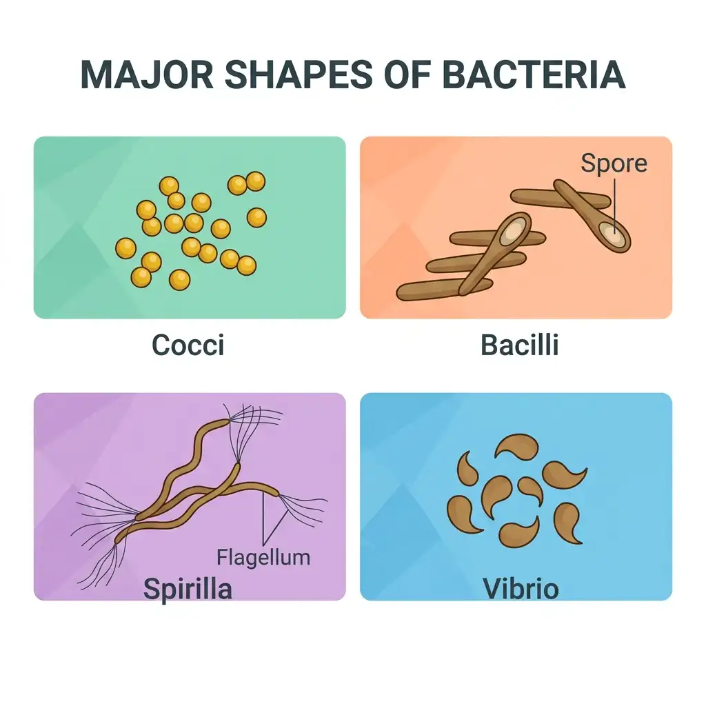
Shapes of Bacteria: Shows the four basic categories of bacteria classified by their shape: Coccus (spherical), Bacillus (rod-shaped), Spirillum (spiral), and Vibrio (comma-shaped).
🔬 Detailed Anatomical Description
Bacteria are microscopic prokaryotic organisms classified into four categories based on their shapes: (1) Cocci (spherical, e.g., Streptococcus); (2) Bacilli (rod-shaped, e.g., Lactobacillus, some of which form highly resistant resting spores); (3) Spirilla (spiral or helical, e.g., Spirillum, containing thin flagella for movement); (4) Vibrio (comma-shaped curved rods, e.g., Vibrio cholerae). This morphological classification is fundamental for microbiology studies.
C BACTERIAL NUTRITION
| MODE | TYPE | DETAIL |
|---|---|---|
| Autotrophic | Photosynthetic autotrophs | Synthesise food using light (e.g., Cyanobacteria) |
| Autotrophic | Chemosynthetic autotrophs | Oxidise inorganic substances (nitrates, nitrites, ammonia); use released energy for ATP; great role in recycling nutrients (N, P, Fe, S) |
| Heterotrophic | — | Depend on other organisms or dead organic matter; most abundant in nature ⭐⭐ |
NEET 2022 / repeated: Heterotrophic bacteria are most abundant in nature. Bacteria show the most extensive METABOLIC diversity (not habitat diversity — that is Fungi).
USEFUL ROLES OF HETEROTROPHIC BACTERIA
- Important decomposers
- Making curd from milk
- Production of antibiotics
- Fixing nitrogen in legume roots
DISEASES CAUSED BY BACTERIA
- Cholera, Typhoid, Tetanus, Citrus canker
BACTERIAL REPRODUCTION
- Mainly by fission (binary fission)
- Under unfavourable conditions → produce spores
- Sexual reproduction: primitive type of DNA transfer from one bacterium to another
A Dividing Bacterium
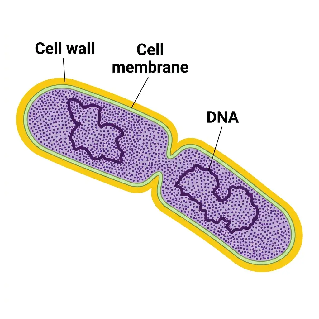
Bacterial Division: Demonstrates binary fission where a single bacterial cell copies its DNA and splits into two identical cells.
🔬 Detailed Anatomical Description
Bacteria reproduce primarily by asexual reproduction via a process called binary fission. During division: (1) The bacterial cell duplicates its single, circular double-stranded DNA molecule; (2) The cell wall and plasma membrane pinch inward along the center, forming a transverse septum; (3) The cytoplasm splits equally, resulting in the separation of two identical daughter cells, each containing a complete copy of the chromosome.
D ARCHAEBACTERIA
| FEATURE | DETAIL |
|---|---|
| Special feature | Live in most harsh habitats |
| Cell wall | Different cell wall structure from other bacteria → responsible for survival in extreme conditions ⭐⭐ |
| Cell membrane | Branched-chain lipids (differ from eubacteria) |
THREE TYPES OF ARCHAEBACTERIA
| TYPE | HABITAT | KEY DETAIL |
|---|---|---|
| Halophiles | Extreme salty areas | — |
| Thermoacidophiles | Hot springs | — |
| Methanogens | Marshy areas; gut of ruminant animals (cows, buffaloes) | Responsible for methane (biogas) production from dung ⭐ |
NEET 2014, 2015, 2016, 2017, 2022: Archaebacteria differ from other bacteria in having different cell wall structure. Methanogens → present in gut of ruminants → responsible for biogas production. Halophiles → found in extreme saline conditions. Thermoacidophiles → hot springs.
E EUBACTERIA ('TRUE BACTERIA')
- Characterised by rigid cell wall
- If motile → have a flagellum
CYANOBACTERIA (BLUE-GREEN ALGAE)
| FEATURE | DETAIL |
|---|---|
| Pigment | Have chlorophyll a (similar to green plants) |
| Nutrition | Photosynthetic autotrophs (oxygenic photosynthesis) |
| Forms | Unicellular, colonial or filamentous |
| Habitat | Freshwater, marine or terrestrial |
| Colony | Generally surrounded by gelatinous sheath |
| Blooms | Often form blooms in polluted water bodies |
| Nitrogen fixation | Some can fix atmospheric nitrogen in specialised cells called heterocysts ⭐⭐ |
| Examples | Nostoc, Anabaena ⭐ |
NEET 2010, 2012, 2018: Nitrogen-fixing cyanobacteria with heterocysts → Nostoc, Anabaena. They perform OXYGENIC photosynthesis (contrast with green sulphur bacteria which perform ANOXYGENIC photosynthesis).
Filamentous Algae - Nostoc
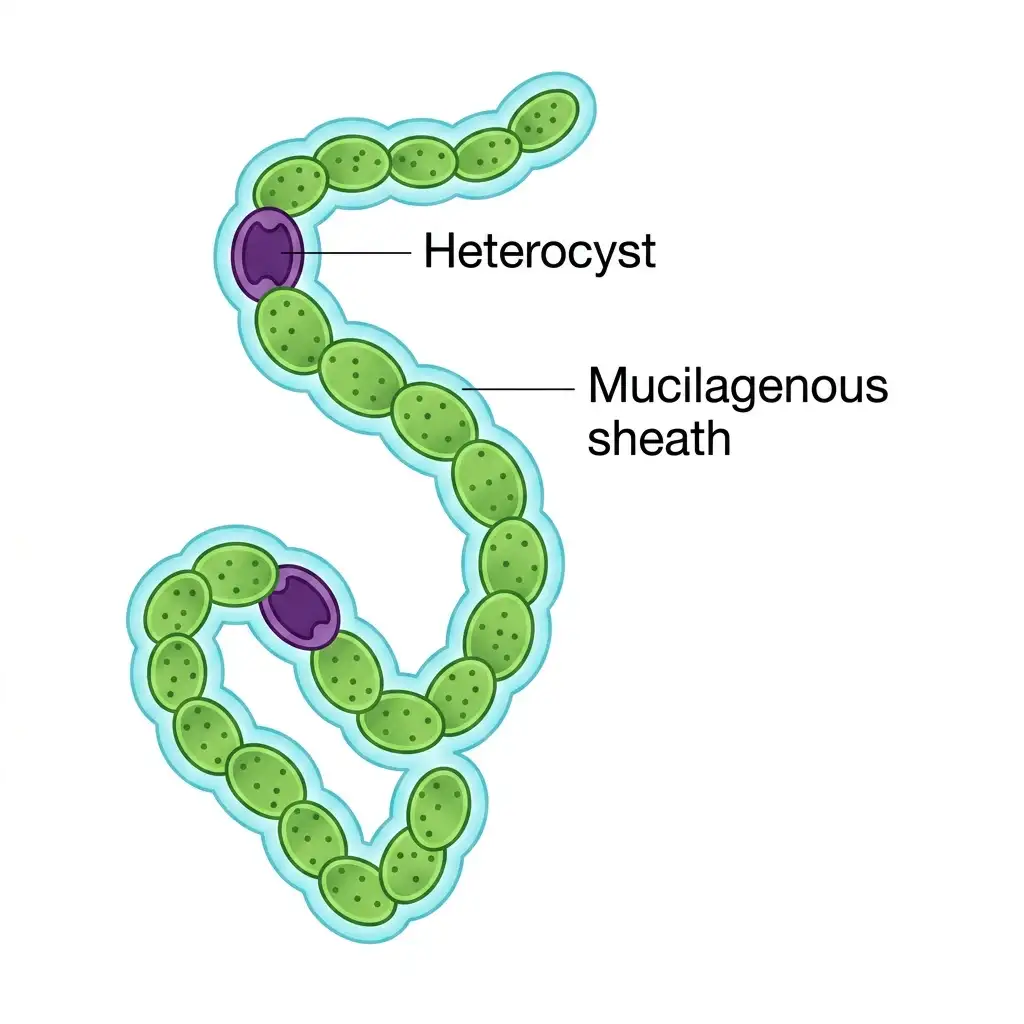
Structure of Nostoc: A multicellular cyanobacteria colony with specialized cells (heterocysts) for nitrogen fixation.
🔬 Detailed Anatomical Description
Nostoc is a genus of filamentous blue-green algae (cyanobacteria) found in terrestrial and aquatic environments. They form macroscopic colonies composed of chains of vegetative bead-like cells wrapped in a protective, gelatinous mucilagenous sheath. Within these filaments, large, specialized thick-walled cells called heterocysts occur. Heterocysts contain the nitrogenase enzyme complex, allowing Nostoc to fix atmospheric nitrogen in anaerobic conditions.
F MYCOPLASMA
| FEATURE | DETAIL |
|---|---|
| Cell type | Prokaryotic |
| Cell wall | Completely LACK cell wall ⭐⭐ |
| Size | Smallest living cells known ⭐⭐ (~0.3 µm) |
| Filter passage | Can pass through 1 micron filter |
| Oxygen | Can survive without oxygen (anaerobic) ⭐ |
| Pathogenicity | Pathogenic to both plants and animals |
NEET 2015, 2017, 2022: Mycoplasma = smallest living cells + completely lack cell wall + can survive without oxygen. Mycoplasma are PROKARYOTIC (Kingdom Monera) — don't confuse with protists!
III. KINGDOM PROTISTA
A GENERAL FEATURES
| FEATURE | DETAIL |
|---|---|
| Cell type | Eukaryotic |
| Organisation | All single-celled (unicellular) ⭐ |
| Habitat | Primarily aquatic |
| Boundaries | Not well defined — shows connections to Plantae, Animalia & Fungi |
| Cell structure | Well-defined nucleus + other membrane-bound organelles |
| Locomotion | Some have flagella or cilia |
| Reproduction | Both asexual and sexual (cell fusion + zygote formation) |
B CHRYSOPHYTES (DIATOMS & GOLDEN ALGAE)
| FEATURE | DETAIL |
|---|---|
| Habitat | Fresh water and marine environments |
| Size | Microscopic |
| Movement | Float passively in water currents (plankton) |
| Nutrition | Most are photosynthetic |
| Cell wall | Two thin overlapping shells fitting together like a SOAP BOX ⭐ |
| Wall composition | Embedded with SILICA → walls are indestructible ⭐ |
| Deposits | Accumulation over billions of years = 'Diatomaceous earth' |
| Uses | Polishing, filtration of oils and syrups |
| Ecological role | Chief producers in the oceans ⭐⭐ |
NEET 2015, 2016, 2018: Diatom cell walls = two overlapping shells (soap box) embedded with silica. Diatoms are chief producers in the oceans. Form diatomaceous earth.
C DINOFLAGELLATES (FIRE ALGAE)
| FEATURE | DETAIL |
|---|---|
| Habitat | Mostly marine |
| Nutrition | Photosynthetic |
| Colour | Yellow, green, brown, blue or red |
| Cell wall | Stiff cellulose plates on outer surface |
| Flagella | Two: one longitudinal, one transverse |
| Red tides | Red dinoflagellates (e.g., Gonyaulax) → rapid multiplication → sea appears red ⭐ |
| Toxins | Released toxins may kill marine animals |
Red tides caused by rapid multiplication of red dinoflagellates (e.g., Gonyaulax). They release toxins. Red Sea colouration is due to Trichodesmium erythraeum (a cyanobacterium) — NOT Gonyaulax.
D EUGLENOIDS
| FEATURE | DETAIL |
|---|---|
| Habitat | Majority freshwater; stagnant water |
| Cell wall | Absent — instead have PELLICLE (protein-rich layer) ⭐ → flexible body |
| Flagella | Two: one short, one long |
| Nutrition | MIXOTROPHIC ⭐ — photosynthetic in sunlight; heterotrophic when deprived |
| Pigments | Identical to those in higher plants |
| Example | Euglena |
| Significance | Connecting link between plants and animals ⭐ |
Euglena has NO cell wall — instead has PELLICLE (protein-rich flexible layer). Mixotrophic (photosynthetic in light, heterotrophic in dark). Connecting link between plants and animals.
E SLIME MOULDS
| FEATURE | DETAIL |
|---|---|
| Nutrition | Saprophytic protists |
| Movement | Body moves along decaying twigs & leaves, engulfing organic material |
| Favourable conditions | Form aggregation called plasmodium (may grow & spread over several feet) |
| Unfavourable conditions | Plasmodium → fruiting bodies bearing spores at tips |
| Spore walls | Possess true walls |
| Spore resistance | Extremely resistant; survive many years |
| Spore dispersal | By air currents |
F PROTOZOANS
Believed to be primitive relatives of animals; all are heterotrophic (predators or parasites)
| GROUP | LOCOMOTION | FEATURES | EXAMPLE |
|---|---|---|---|
| Amoeboid protozoans | Pseudopodia (false feet) | Fresh/sea water, moist soil; marine forms have silica shells; some parasitic | Amoeba, Entamoeba ⭐ |
| Flagellated protozoans | Flagella | Free-living or parasitic; cause sleeping sickness | Trypanosoma ⭐ |
| Ciliated protozoans | Thousands of cilia | Aquatic; cavity (gullet); coordinated cilia; TWO types of nuclei (macro + micronucleus) | Paramoecium ⭐ |
| Sporozoans | No locomotory structures | Infectious spore-like stage in life cycle | Plasmodium (malarial parasite) ⭐ |
NEET 2018, 2024: Paramoecium has TWO types of nuclei (macronucleus + micronucleus). Sporozoans (e.g., Plasmodium) = parasitic; do NOT have locomotory structures.
Different Types of Protists
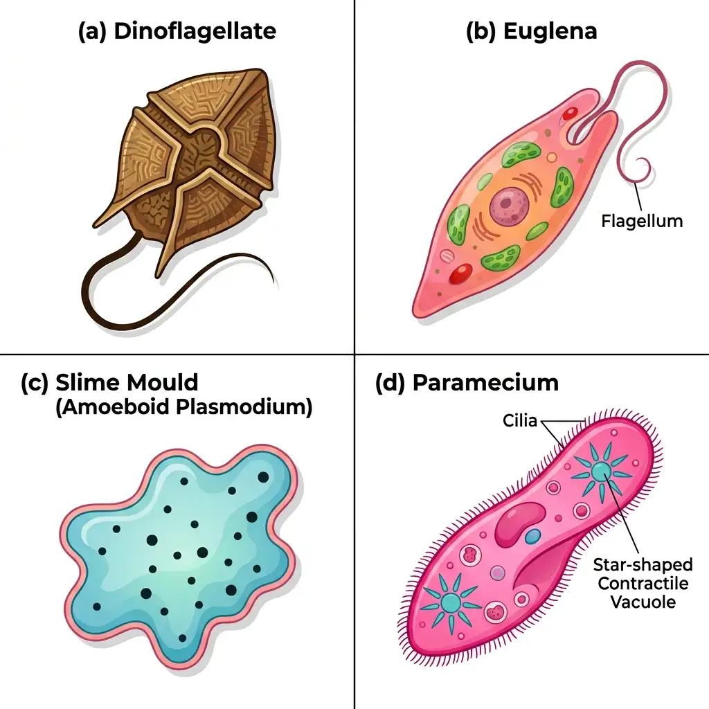
Diversity of Kingdom Protista: Compares representative members including dinoflagellates, euglenoids (Euglena), slime moulds, and ciliates (Paramecium).
🔬 Detailed Anatomical Description
Kingdom Protista comprises single-celled eukaryotic organisms. This figure illustrates key representatives: (a) Dinoflagellates: Armored cells with cellulose plates and two flagella causing red tides; (b) Euglena: Photosynthetic mixotroph with a protein-rich pellicle instead of a cell wall; (c) Slime mould: Saprophytic protist forming amoeboid plasmodium structures; (d) Paramecium: Ciliated protozoan containing a cytostome mouth, macronucleus, and contractile vacuoles.
IV. KINGDOM FUNGI
A GENERAL FEATURES
| FEATURE | DETAIL |
|---|---|
| Nutrition | Heterotrophic — unique kingdom of heterotrophic organisms |
| Cell wall | Composed of CHITIN and polysaccharides ⭐⭐ (chitin = homopolymer) |
| Body | Filamentous (exception: yeasts = unicellular) ⭐ |
| Structure | Long, slender thread-like hyphae → network = mycelium ⭐ |
| Hyphae types | Coenocytic (aseptate, continuous, multinucleated) OR Septate (with cross walls) |
| Habitat | Cosmopolitan; prefer warm & humid |
| Diversity | Great diversity in morphology and habitat (extensive HABITAT diversity) |
MODES OF NUTRITION
| MODE | DETAIL |
|---|---|
| Saprophytes | Absorb soluble organic matter from dead substrates (most fungi) |
| Parasites | Depend on living plants and animals |
| Symbionts | With algae → Lichens; With roots of higher plants → Mycorrhiza |
NEET 2022: Fungi show extensive HABITAT diversity (not metabolic diversity — that is bacteria). All fungi are heterotrophic (saprophytic, parasitic or symbiotic). Cell wall = CHITIN (not cellulose).
B REPRODUCTION IN FUNGI
SEXUAL REPRODUCTION — THREE STEPS
| STEP | PROCESS |
|---|---|
| 1. Plasmogamy | Fusion of protoplasms between two gametes |
| 2. Karyogamy | Fusion of two nuclei |
| 3. Meiosis | In zygote → produces haploid spores |
NEET 2021: Sexual cycle in fungi = Plasmogamy → Karyogamy → Meiosis. Dikaryophase found in Ascomycetes and Basidiomycetes ONLY.
C BASIS FOR CLASSIFICATION OF FUNGI INTO CLASSES
| CRITERION | DETAIL |
|---|---|
| 1. Morphology of mycelium | Septate vs aseptate/coenocytic |
| 2. Mode of spore formation | Type of asexual and sexual spores |
| 3. Fruiting bodies | Structure of fruiting bodies |
NEET 2024: Classification of fungi into classes is based on mycelium morphology, spore formation & fruiting bodies. Mode of nutrition is NOT a criterion (all fungi are heterotrophic — nutrition doesn't distinguish classes)!
D FOUR CLASSES OF FUNGI — MASTER COMPARISON TABLE
| FEATURE | PHYCOMYCETES | ASCOMYCETES (SAC FUNGI) | BASIDIOMYCETES (CLUB FUNGI) | DEUTEROMYCETES (IMPERFECT FUNGI) |
|---|---|---|---|---|
| Mycelium | Aseptate & coenocytic ⭐ | Septate & branched | Septate & branched | Septate & branched |
| Habitat | Aquatic, decaying wood, moist, obligate parasites | Saprophytic, decomposers, parasitic, coprophilous | Soil, logs, tree stumps, parasites on living plants | Saprophytes, parasites, decomposers (mineral cycling) ⭐ |
| Asexual spores | Zoospores (motile) or Aplanospores (non-motile); endogenous | Conidia (exogenously on conidiophores) ⭐ | Generally not found; vegetative repro by fragmentation | Conidia only |
| Sexual spores | Zygospore | Ascospores (endogenously in asci; ascocarps) ⭐ | Basidiospores (exogenously on basidium; basidiocarps) ⭐ | Sexual repro absent / unknown ⭐ |
| Dikaryophase | ❌ | ✅ Yes | ✅ Yes | — |
| Examples | Mucor, Rhizopus (bread mould) ⭐, Albugo (parasitic on mustard) | Aspergillus, Claviceps, Neurospora ⭐, Penicillium, Yeast, Morels, Truffles | Agaricus (mushroom) ⭐, Ustilago (smut) ⭐, Puccinia (rust) ⭐ | Alternaria, Colletotrichum, Trichoderma ⭐ |
NEET 2024: Rhizopus (bread mould) = Phycomycetes; mycelium is aseptate and coenocytic. In Ascomycetes, Basidiomycetes, and Deuteromycetes → mycelium is septate. Only Phycomycetes is aseptate.
Different Types of Fungi
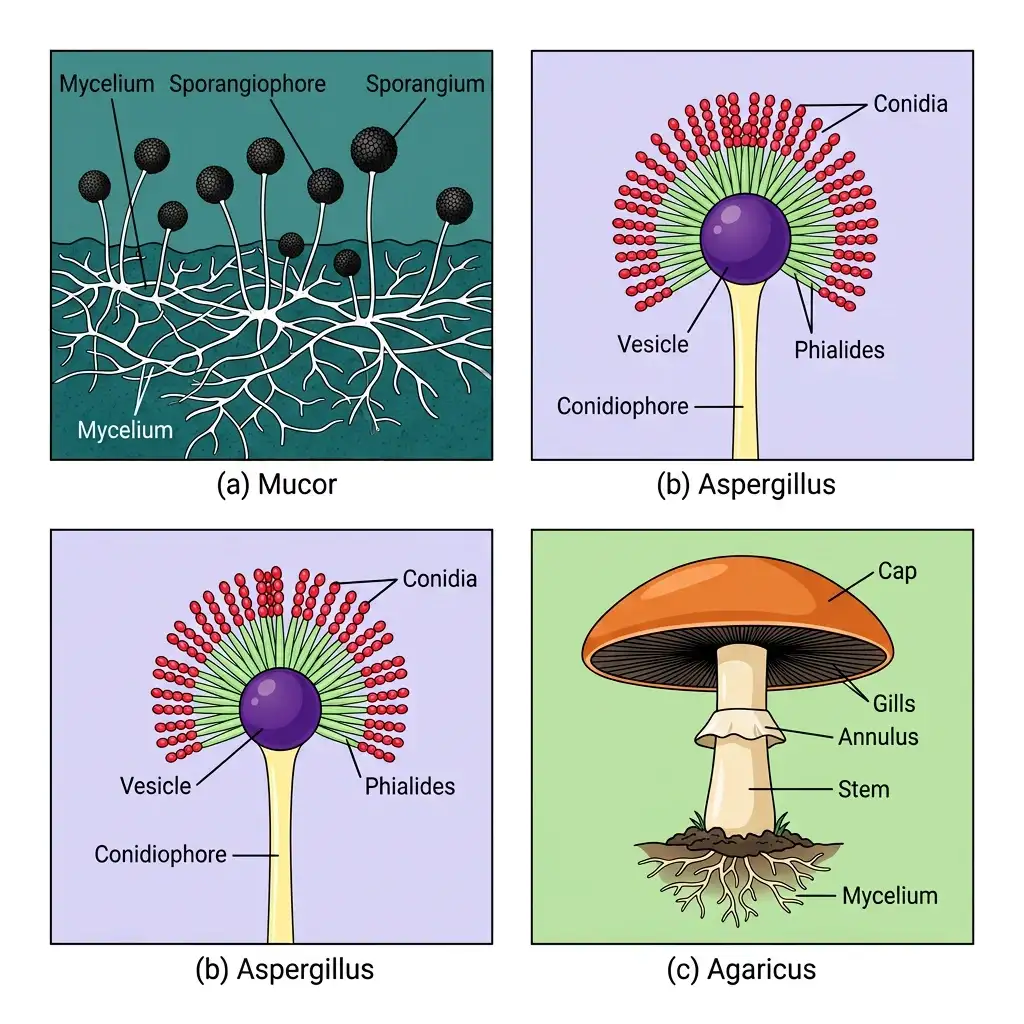
Kingdom Fungi Representatives: Compares filamentous moulds (Mucor, Aspergillus) with multicellular basidiomycetes (Agaricus field mushroom).
🔬 Detailed Anatomical Description
Kingdom Fungi consists of heterotrophic, eukaryotic organisms with chitinous cell walls. This diagram compares: (a) Mucor: A typical pin mould with branching non-septate coenocytic hyphae ending in rounded black sporangia; (b) Aspergillus: A septate ascomycete with conidiophores releasing chains of asexual conidiospores; (c) Agaricus: The common field mushroom displaying a subterranean mycelium, an aerial stalk (stipe), and a cap (pileus) with gills.
KEY EXAMPLES — DETAILED
YEAST (Saccharomyces cerevisiae)
| FEATURE | DETAIL |
|---|---|
| Classification | Ascomycetes |
| Also known as | Baker's yeast and Brewer's yeast |
| Unique feature | Only unicellular and non-filamentous fungus ⭐ |
| Uses | Making bread and beer |
NEUROSPORA
| FEATURE | DETAIL |
|---|---|
| Classification | Ascomycetes |
| Use | Used extensively in biochemical and genetic work |
| Known as | 'Drosophila of plant kingdom' ⭐ |
EDIBLE FUNGI
| FUNGI | TYPE | CLASS |
|---|---|---|
| Morels | Edible delicacy | Ascomycetes |
| Truffles | Edible delicacy | Ascomycetes |
| Agaricus (Mushroom) | Commonly eaten | Basidiomycetes |
DISEASE-CAUSING FUNGI
| DISEASE | FUNGUS | CLASS |
|---|---|---|
| Early blight of potato | Alternaria | Deuteromycetes |
| Late blight of potato | Phytophthora infestans | Phycomycetes (Oomycete) |
| Wheat rust | Puccinia | Basidiomycetes |
| Smut | Ustilago | Basidiomycetes |
| White spots on mustard | Albugo | Phycomycetes |
SPECIAL EXAMPLES
| ORGANISM | CLASS | SPECIAL FEATURE |
|---|---|---|
| Claviceps | Ascomycetes | Source of LSD (ergot alkaloids) |
| Penicillium | Ascomycetes | Source of antibiotics |
| Amanita muscaria | Basidiomycetes | Contains hallucinogens |
Only UNICELLULAR fungus = Yeast (Saccharomyces cerevisiae) — belongs to Ascomycetes. Neurospora = 'Drosophila of the plant kingdom' (Ascomycetes). Agaricus (mushroom), Ustilago (smut), Puccinia (rust) = Basidiomycetes. Penicillium, Aspergillus, Claviceps, Neurospora, Yeast, Morels, Truffles = Ascomycetes.
V. KINGDOM PLANTAE (BRIEF)
| FEATURE | DETAIL |
|---|---|
| Includes | All eukaryotic chlorophyll-containing organisms |
| Cell wall | Cellulose |
| Cell structure | Eukaryotic with prominent chloroplasts |
| Partially heterotrophic | Insectivorous: Bladderwort, Venus fly trap; Parasite: Cuscuta |
| Groups | Algae, Bryophytes, Pteridophytes, Gymnosperms, Angiosperms |
| Life cycle | Alternation of generations — diploid sporophytic phase alternates with haploid gametophytic phase ⭐ |
VI. KINGDOM ANIMALIA (BRIEF)
| FEATURE | DETAIL |
|---|---|
| Cell type | Eukaryotic |
| Cell wall | ABSENT ⭐⭐ |
| Nutrition | Heterotrophic (holozoic — ingestion of food) |
| Organisation | Multicellular |
| Food reserves | Stored as glycogen or fat |
| Growth | Definite pattern; adults have definite shape & size |
| Higher forms | Elaborate sensory and neuromotor mechanism |
| Locomotion | Most are capable |
| Reproduction | Sexual — copulation → embryological development |
VII. VIRUSES
A HISTORY
| SCIENTIST | YEAR | CONTRIBUTION |
|---|---|---|
| Dmitri Ivanowsky | 1892 | Recognised microbes causing mosaic disease of tobacco; smaller than bacteria (passed through filters) |
| M.W. Beijerinck | 1898 | Demonstrated infection from extract; named 'virus'; called the fluid 'Contagium vivum fluidum' ⭐⭐ |
| W.M. Stanley | 1935 | Showed viruses could be crystallised; crystals consist largely of proteins ⭐ |
NEET 2015: Beijerinck (1898) = 'Contagium vivum fluidum'. 'Virus' means venom or poisonous fluid.
B GENERAL FEATURES
| FEATURE | DETAIL |
|---|---|
| Nature | Non-cellular organisms; not truly living |
| Outside host | Inert crystalline structure |
| Inside host | Take over host cell machinery → replicate → kill host |
| Parasitism | Obligate parasites ⭐⭐ |
| Composition | Nucleoprotein (protein + nucleic acid) |
| Genetic material | Either RNA or DNA — NEVER BOTH ⭐⭐ |
| Infectious part | The genetic material is infectious |
C VIRUS STRUCTURE
| COMPONENT | DETAIL |
|---|---|
| Capsid | Protein coat protecting nucleic acid |
| Capsomeres | Small subunits making up the capsid ⭐ |
| Arrangement | Capsomeres arranged in helical or polyhedral geometric forms ⭐ |
D GENETIC MATERIAL IN DIFFERENT VIRUSES
| VIRUS TYPE | GENETIC MATERIAL |
|---|---|
| Plant viruses | Single-stranded RNA (ssRNA) |
| Animal viruses | ss or ds RNA OR ds DNA |
| Bacteriophages | Usually double-stranded DNA (dsDNA) |
NEET 2014, 2019: Viruses have either DNA or RNA (never both). Genetic material is the infectious part. TMV = coiled RNA + capsomeres (single-stranded RNA + protein coat capsid made of capsomeres).
TMV and Bacteriophage
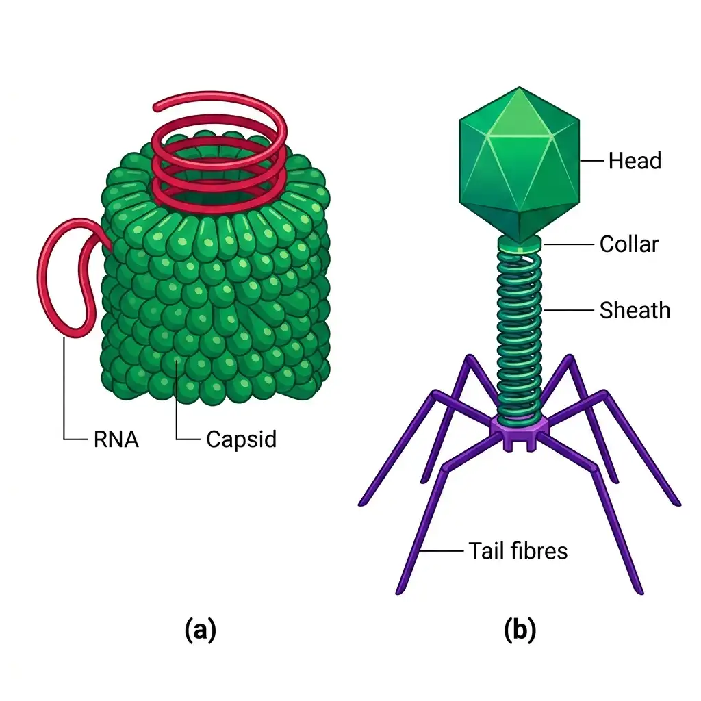
Viral Morphology: Illustrates non-cellular infectious agents, featuring helical plant TMV and complex bacterial T4 phage with head and tail fibers.
🔬 Detailed Anatomical Description
Viruses are non-cellular, obligate intracellular parasites consisting of a genetic core wrapped in a protein coat (capsid). This diagram details: (a) Tobacco Mosaic Virus (TMV): A helical plant virus containing single-stranded RNA wrapped in repeating protein capsomeres; (b) T4 Bacteriophage: A complex virus that infects bacteria, comprising an icosahedral head capsule containing double-stranded DNA, a collar neck, a contractile sheath, and tail fibers.
E DISEASES CAUSED BY VIRUSES
IN HUMANS:
- Mumps, Smallpox, Herpes, Influenza, AIDS
IN PLANTS — SYMPTOMS:
- Mosaic formation, Leaf rolling & curling, Yellowing & vein clearing, Dwarfing & stunted growth
VIII. VIROIDS
| FEATURE | DETAIL |
|---|---|
| Discovered by | T.O. Diener in 1971 ⭐ |
| Size | Smaller than viruses |
| Composition | Free RNA of low molecular weight ⭐ |
| Protein coat | ABSENT ⭐⭐ |
| Disease caused | Potato spindle tuber disease ⭐ |
NEET 2016, 2017: Viroids discovered by T.O. Diener (1971) (NOT Ivanowsky). Viroids LACK protein coat. Composition: FREE RNA (low molecular weight). Smaller than viruses. Cause potato spindle tuber disease.
IX. PRIONS
| FEATURE | DETAIL |
|---|---|
| Composition | Abnormally folded proteins ⭐ |
| Size | Similar in size to viruses |
| Nature | Infectious neurological disease agents |
DISEASES CAUSED BY PRIONS
| DISEASE | AFFECTED |
|---|---|
| BSE (Bovine Spongiform Encephalopathy) = Mad Cow Disease | Cattle ⭐ |
| CJD (Creutzfeldt-Jakob Disease) | Humans (analogous variant) ⭐ |
RE-NEET 2026 + NEET 2019, 2020, 2022: Prions = abnormally folded proteins (no nucleic acid). BSE (mad cow disease) in cattle; CJD in humans. NOT caused by Mycoplasma, viroids or Aspergillus.
X. LICHENS
| FEATURE | DETAIL |
|---|---|
| Nature | Symbiotic association (mutually useful) between algae and fungi |
| Algal component | Phycobiont (autotrophic — prepares food for fungi) |
| Fungal component | Mycobiont (heterotrophic — provides shelter, absorbs minerals & water) |
| Pollution indicator | Lichens do NOT grow in polluted areas ⭐⭐ (especially indicate SO₂ pollution) |
| Appearance | So closely associated that they appear as a single organism in nature |
NEET 2012, 2014, 2015, 2019, RE-NEET 2026: Lichens = symbiotic association between algae (phycobiont — autotrophic) and fungi (mycobiont — heterotrophic) (not mycorrhiza or sponges or chrysophytes). They are very good pollution indicators (do not grow in polluted areas; specifically indicate SO₂ pollution). Luxuriant growth of lichens = location is NOT polluted.
XI. RAPID REVISION — ALL EXAMPLES TABLE
| CATEGORY | EXAMPLES |
|---|---|
| Archaebacteria — Halophiles | Extreme salty areas |
| Archaebacteria — Thermoacidophiles | Hot springs |
| Archaebacteria — Methanogens | Marshy areas; gut of ruminants; produce methane/biogas |
| Cyanobacteria with heterocysts | Nostoc, Anabaena (nitrogen fixation) |
| Smallest living cells | Mycoplasma |
| Chrysophytes — Chief ocean producers | Diatoms (silica walls, soap-box shape) |
| Dinoflagellates — Red tides | Gonyaulax |
| Red Sea colouration | Trichodesmium erythraeum (cyanobacterium) |
| Euglenoids | Euglena (mixotrophic; pellicle; connecting link) |
| Slime moulds | Saprophytic; plasmodium; fruiting bodies with spores |
| Amoeboid protozoans | Amoeba, Entamoeba |
| Flagellated protozoans | Trypanosoma (sleeping sickness) |
| Ciliated protozoans | Paramoecium (two nuclei; gullet) |
| Sporozoans | Plasmodium (no locomotory structures) |
| Phycomycetes | Mucor, Rhizopus (bread mould), Albugo |
| Ascomycetes (Sac fungi) | Aspergillus, Claviceps, Neurospora, Penicillium, Yeast, Morels, Truffles |
| Basidiomycetes (Club fungi) | Agaricus (mushroom), Ustilago (smut), Puccinia (rust), Amanita muscaria |
| Deuteromycetes (Imperfect) | Alternaria, Colletotrichum, Trichoderma |
| Insectivorous plants | Bladderwort, Venus fly trap |
| Parasitic plant | Cuscuta |
| Virus — TMV | ssRNA |
| Virus — Bacteriophage | dsDNA |
| Viroid | Potato spindle tuber disease; free RNA; no protein coat |
| Prion diseases | BSE (cattle); CJD (humans) |
| Lichens | Algae (phycobiont) + Fungi (mycobiont); pollution indicator |
NEET 2025 / 2026 / RE-NEET 2026 ONE-LINERS (HIGH-YIELD TRAPS) ⚡
- Mad cow disease (BSE) = Prions (abnormally folded proteins)
- Symbiotic fungi + algae = Lichens
- Not a prokaryote = Fungi
- Bacterial shapes: Spherical = Cocci | Rod = Bacilli | Comma = Vibrio | Spirillum = Spirilla
- Only non-membrane bound organelle in BOTH prokaryotes & eukaryotes = Ribosomes (70S in prok; 80S cytoplasmic + 70S in organelles in euk)
- Viroid = free RNA, no protein coat (Diener 1971)
- Archaebacteria cell wall different (survive extremes)
- Mycoplasma = smallest cells + no cell wall
- Diatoms = chief ocean producers + silica soap-box shells
- Red tides = Gonyaulax (dinoflagellate)
- Dikaryophase = Ascomycetes & Basidiomycetes only
- Phycomycetes mycelium = aseptate/coenocytic
XII. COMPARISON TABLE — VIRUS VS VIROID VS PRION
| FEATURE | VIRUS | VIROID | PRION |
|---|---|---|---|
| Discovered by | Ivanowsky (1892); Beijerinck (1898); Stanley (1935) | T.O. Diener (1971) | Modern medicine |
| Size | Small | Smaller than viruses | Similar to viruses |
| Composition | Nucleic acid + Protein coat (nucleoprotein) | Only RNA (free RNA, low MW) | Only protein (abnormally folded) |
| Protein coat | Present (capsid with capsomeres) | ABSENT ⭐ | Entire agent is protein |
| Nucleic acid | DNA or RNA (never both) | Only RNA | Absent |
| Nature | Obligate parasites; non-cellular | Infectious agent | Infectious agent |
| Disease examples | Mumps, AIDS, Mosaic disease | Potato spindle tuber disease | BSE (cattle), CJD (humans) |
XIII. COMMON EXAM TRAPS — QUICK REFERENCE
| TRAP / QUESTION | CORRECT ANSWER |
|---|---|
| Most extensive metabolic diversity? | Bacteria (Monera) — NOT fungi |
| Extensive habitat diversity? | Fungi — NOT bacteria |
| Most abundant bacteria in nature? | Heterotrophic bacteria |
| Smallest living cells? | Mycoplasma (not viruses — viruses are not cells) |
| Mycoplasma has cell wall? | No — completely lacks cell wall |
| Chief producers in oceans? | Diatoms (Chrysophytes) |
| Red tides caused by? | Gonyaulax (red dinoflagellate) |
| Red Sea coloured by? | Trichodesmium erythraeum (cyanobacterium) — NOT Gonyaulax |
| Euglena — plant or animal? | Neither — connecting link (Protista; mixotrophic) |
| Euglena has cell wall? | No — has pellicle (protein-rich layer) |
| Only unicellular fungus? | Yeast (Saccharomyces cerevisiae) |
| Yeast belongs to which class? | Ascomycetes |
| Neurospora = 'Drosophila of'? | Plant kingdom |
| Phycomycetes mycelium? | Aseptate & coenocytic |
| Dikaryophase found in? | Ascomycetes & Basidiomycetes only |
| Imperfect fungi? | Deuteromycetes (sexual repro absent/unknown) |
| Deuteromycetes when sexual stage found? | Moved to Ascomycetes or Basidiomycetes |
| Conidia produced exogenously on conidiophores? | Ascomycetes (asexual spores) |
| Basidiospores produced exogenously? | On basidium (Basidiomycetes) |
| Ascospores produced endogenously? | In asci (Ascomycetes) |
| Mode of nutrition as basis for classifying fungi into classes? | NO — criterion is mycelium morphology, spore formation, fruiting bodies |
| Virus has both DNA and RNA? | No — either DNA or RNA, never both |
| Viroid discovered by? | T.O. Diener (1971) — NOT Ivanowsky |
| Viroid has protein coat? | No — free RNA only |
| Prion composition? | Abnormally folded protein (no nucleic acid) |
| Lichens grow in polluted areas? | No — pollution indicators; do not grow in polluted areas |
| Algal component of lichen? | Phycobiont (autotrophic) |
| Fungal component of lichen? | Mycobiont (heterotrophic) |
| Alternation of generations in? | Kingdom Plantae |
| Animals store food as? | Glycogen or fat (not starch) |
| Animals lack? | Cell wall |
- RE-NEET 2026 trap: Mad cow disease = prions (not Mycoplasma/viroids/Aspergillus).
- RE-NEET 2026 trap: Lichens = fungi + algae symbiotic association.
- RE-NEET 2026 trap: Fungi is NOT prokaryote.
- RE-NEET 2026 trap: Correct bacterial shape matching (Cocci, Bacilli, Vibrio, Spirilla).
- NEET 2026 trap: Ribosomes = common non-membrane organelle in prokaryotes and eukaryotes.
- Reinforce: Prions = abnormally folded proteins (no nucleic acid).
- Reinforce: Lichens do not grow in polluted areas (SO₂ indicators).