I. INTRODUCTION
| FEATURE | DETAIL |
|---|---|
| Need for circulation | Cells need continuous supply of nutrients, O₂, and other essential substances and removal of wastes/harmful substances ⭐ |
| Simple organisms | Sponges and coelenterates circulate water through body cavities for exchange ⭐ |
| More complex organisms | Use special body fluids for transport ⭐ |
| Main body fluids | Blood and lymph ⭐⭐ |
| Blood | Most commonly used body fluid in higher organisms including humans ⭐ |
II. BLOOD
| FEATURE | DETAIL |
|---|---|
| Blood | Special connective tissue ⭐⭐⭐ |
| Components | Fluid matrix = plasma + formed elements ⭐⭐⭐ |
NEET 2015: Blood = special connective tissue composed of plasma and formed elements.
III. PLASMA
| FEATURE | DETAIL |
|---|---|
| Nature | Straw-coloured, viscous fluid ⭐⭐ |
| Percentage of blood | About 55% of blood ⭐⭐⭐ |
| Water content | 90–92% ⭐⭐ |
| Protein content | 6–8% ⭐⭐ |
Major Plasma Proteins ⭐⭐⭐
| PLASMA PROTEIN | FUNCTION |
|---|---|
| Fibrinogen | Coagulation / clotting of blood ⭐⭐⭐ |
| Globulins | Defense mechanism / antibodies ⭐⭐⭐ |
| Albumins | Osmotic balance ⭐⭐⭐ |
NEET 2018: Know functions of fibrinogen, globulins, albumins. NEET 2015: Antibody deficiency can be checked by serum globulins.
Other Plasma Components ⭐⭐
| COMPONENT | DETAIL |
|---|---|
| Minerals present | Na⁺, Ca²⁺, Mg²⁺, HCO₃⁻, Cl⁻ etc. ⭐ |
| Nutrients present | Glucose, amino acids, lipids etc. in transit ⭐ |
| Clotting factors | Present in inactive form in plasma ⭐⭐ |
Serum ⭐⭐⭐
| FEATURE | DETAIL |
|---|---|
| Serum | Plasma without clotting factors ⭐⭐⭐ |
NEET 2016: Serum = plasma without clotting factors.
IV. FORMED ELEMENTS
| FORMED ELEMENT | COUNT / FEATURES | KEY FUNCTION |
|---|---|---|
| Erythrocytes (RBCs) | Most abundant; 5–5.5 million/mm³ ⭐⭐⭐ | Transport respiratory gases ⭐⭐ |
| Leucocytes (WBCs) | 6000–8000/mm³; colourless due to lack of Hb ⭐⭐ | Defense / immunity ⭐⭐ |
| Platelets (Thrombocytes) | 1,50,000–3,50,000/mm³ ⭐⭐⭐ | Clotting / coagulation ⭐⭐⭐ |
Formed Elements in Blood
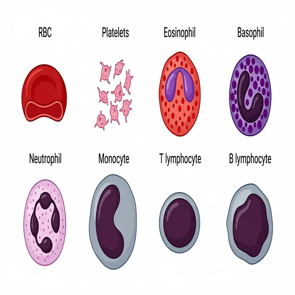
Formed Elements in Blood: Erythrocytes (RBCs), Leucocytes (WBCs), and Platelets constitute the formed elements of blood.
🔬 Detailed Anatomical Description
Blood contains plasma and formed elements. (1) Erythrocytes (RBCs) are bi-concave and lack nuclei, carrying oxygen. (2) Leucocytes (WBCs) include granulocytes (neutrophils, eosinophils, basophils) and agranulocytes (lymphocytes, monocytes) responsible for immune defense. (3) Platelets (thrombocytes) are cell fragments crucial for blood clotting.
V. ERYTHROCYTES (RBCS)
| FEATURE | DETAIL |
|---|---|
| Other name | Red blood cells (RBCs) / Erythrocytes ⭐ |
| Most abundant blood cells | Yes ⭐⭐⭐ |
| Count | 5 to 5.5 million/mm³ ⭐⭐ |
| Site of formation | Red bone marrow ⭐⭐⭐ |
| Shape | Biconcave ⭐⭐ |
| Nucleus | Absent in most mammals / enucleated ⭐⭐⭐ |
| Why enucleated? | To make maximum space available for O₂ transport ⭐⭐ |
| Pigment | Haemoglobin (iron-containing red coloured complex protein) ⭐⭐⭐ |
| Hb level | 12–16 g per 100 mL blood ⭐⭐⭐ |
| Lifespan | 120 days ⭐⭐⭐ |
| Site of destruction | Spleen = graveyard of RBCs ⭐⭐⭐ |
NEET 2017, 2022: Spleen = graveyard of erythrocytes. NEET 2022: Adult RBCs are enucleated. NEET 2012, 2022: RBC lifespan = 120 days.
VI. LEUCOCYTES (WBCS)
A BASIC FACTS
| FEATURE | DETAIL |
|---|---|
| Other name | White blood cells (WBCs) / Leucocytes ⭐ |
| Colour | Colourless due to lack of haemoglobin ⭐⭐ |
| Nucleus | Present (nucleated) ⭐⭐ |
| Count | 6000–8000/mm³ ⭐⭐ |
| Lifespan | Generally short lived ⭐ |
B TWO MAIN CATEGORIES
| CATEGORY | TYPES |
|---|---|
| Granulocytes | Neutrophils, Eosinophils, Basophils ⭐⭐⭐ |
| Agranulocytes | Lymphocytes, Monocytes ⭐⭐⭐ |
NEET 2020: WBC classification — granulocytes and agranulocytes.
C DIFFERENTIAL LEUKOCYTE COUNT
| WBC TYPE | PERCENTAGE | FUNCTION |
|---|---|---|
| Neutrophils | 60–65% ⭐⭐⭐ | Phagocytosis ⭐⭐⭐ |
| Lymphocytes | 20–25% ⭐⭐⭐ | Immune response ⭐⭐⭐ |
| Monocytes | 6–8% ⭐⭐⭐ | Phagocytosis ⭐⭐ |
| Eosinophils | 2–3% ⭐⭐⭐ | Resist infections; associated with allergic reactions ⭐⭐⭐ |
| Basophils | 0.5–1% ⭐⭐⭐ | Inflammatory reactions; secrete histamine, serotonin, heparin ⭐⭐⭐ |
D HIGH-YIELD WBC FACTS
| FEATURE | DETAIL |
|---|---|
| Most abundant WBC | Neutrophils ⭐⭐⭐ |
| Least abundant WBC | Basophils ⭐⭐⭐ |
| Phagocytic cells | Neutrophils and Monocytes ⭐⭐⭐ |
| Immune response | B and T lymphocytes responsible ⭐⭐⭐ |
| Basophils secrete | Histamine, serotonin, heparin ⭐⭐⭐ |
| Basophils involved in | Inflammatory reactions ⭐⭐⭐ |
| Eosinophils | Resist infections; associated with allergies ⭐⭐ |
| Neutrophils | Strictly phagocytic ⭐⭐ |
NEET 2023: Basophils = least abundant; involved in inflammatory reactions; secrete histamine, serotonin, heparin. WBC abundance order = Neutrophils > Lymphocytes > Monocytes > Eosinophils > Basophils.
VII. PLATELETS / THROMBOCYTES
| FEATURE | DETAIL |
|---|---|
| Other name | Thrombocytes / Platelets ⭐⭐ |
| Nature | Cell fragments ⭐⭐⭐ |
| Produced from | Megakaryocytes (special cells in bone marrow) ⭐⭐⭐ |
| Count | 1,50,000–3,50,000/mm³ of blood ⭐⭐⭐ |
| Function | Release substances involved in coagulation/clotting of blood ⭐⭐⭐ |
| Low platelet count | Causes clotting disorders → excessive blood loss ⭐⭐⭐ |
NEET 2016: Platelets are cell fragments from megakaryocytes; low number → clotting disorder.
VIII. BLOOD GROUPS
A ABO GROUPING
| BLOOD GROUP | ANTIGEN ON RBCS | ANTIBODY IN PLASMA | DONOR COMPATIBILITY |
|---|---|---|---|
| A | A | Anti-B | A, O |
| B | B | Anti-A | B, O |
| AB | A and B | Nil | AB, A, B, O |
| O | Nil | Anti-A and Anti-B | O |
NEET 2012, 2014: O group has no antigen on RBCs.
B KEY ABO FACTS
| FEATURE | DETAIL |
|---|---|
| Basis of ABO grouping | Presence/absence of A and B antigens on RBCs ⭐⭐⭐ |
| Plasma contains | Natural antibodies against absent antigens ⭐⭐ |
| Universal donor | O group ⭐⭐⭐ |
| Universal recipient | AB group / AB⁺ in clinical context ⭐⭐⭐ |
| Why AB is universal recipient? | Because plasma has no anti-A or anti-B antibodies ⭐⭐⭐ |
| Wrong transfusion result | Clumping / destruction of RBCs ⭐⭐ |
NEET 2014, 2021: AB group = universal recipient. NEET 2024: A Rh⁻ person can receive only A Rh⁻ and O Rh⁻.
C Rh GROUPING
| FEATURE | DETAIL |
|---|---|
| Rh antigen | Similar to one in Rhesus monkey ⭐ |
| If antigen present | Rh positive (Rh⁺) ⭐⭐⭐ |
| If absent | Rh negative (Rh⁻) ⭐⭐⭐ |
| Rh⁻ person exposed to Rh⁺ blood | Forms antibodies against Rh antigen ⭐⭐ |
| Rh must be matched before transfusion | Yes ⭐⭐⭐ |
D Rh INCOMPATIBILITY
| FEATURE | DETAIL |
|---|---|
| Special case | Rh⁻ mother carrying Rh⁺ foetus ⭐⭐⭐ |
| First pregnancy | Usually normal because foetal and maternal blood separated by placenta ⭐⭐⭐ |
| During first delivery | Small amount of foetal Rh⁺ blood may enter mother → mother forms anti-Rh antibodies ⭐⭐⭐ |
| Second Rh⁺ pregnancy | Maternal antibodies may destroy foetal RBCs ⭐⭐⭐ |
| Result | Severe anaemia and jaundice in baby; may be fatal ⭐⭐⭐ |
| Name of disease | Erythroblastosis foetalis ⭐⭐⭐ |
| Prevention | Give anti-Rh antibodies to mother immediately after first delivery ⭐⭐⭐ |
NEET 2020, 2021, 2026 context / reinforced: Rh incompatibility occurs when an Rh⁻ mother carries an Rh⁺ foetus. First pregnancy usually normal; problems usually appear in the second or subsequent pregnancies leading to erythroblastosis foetalis.
IX. COAGULATION OF BLOOD
A OVERVIEW
| FEATURE | DETAIL |
|---|---|
| Trigger | Injury/trauma stimulates coagulation ⭐⭐ |
| Purpose | Prevent excessive blood loss ⭐⭐⭐ |
| Clot / Coagulum | Formed mainly of network of fibrin threads trapping dead and damaged formed elements ⭐⭐⭐ |
NEET 2022, NEET 2026 / reinforced: Blood clot is formed by a network of fibrin threads (not thrombin threads — TRAP!).
B CLOTTING CASCADE
| CONVERSION | BY |
|---|---|
| Prothrombin (inactive) → Thrombin (active) | Thrombokinase / enzyme complex ⭐⭐⭐ |
| Fibrinogen (inactive) → Fibrin (active) | Thrombin ⭐⭐⭐ |
C IMPORTANT FACTS
| FEATURE | DETAIL |
|---|---|
| Platelets role | Release factors that activate coagulation mechanism ⭐⭐⭐ |
| Tissue factors | Released from injury site can also initiate coagulation ⭐⭐ |
| Calcium ions | Play a very important role in clotting ⭐⭐⭐ |
| Clotting factors in plasma | Present in inactive state ⭐⭐ |
NEET 2021: Thrombin converts fibrinogen into fibrin. Clotting pathway — prothrombin → thrombin → fibrinogen → fibrin.
X. LYMPH / TISSUE FLUID
| FEATURE | DETAIL |
|---|---|
| Tissue fluid / interstitial fluid | Fluid that comes out of capillaries into spaces between tissue cells ⭐⭐⭐ |
| Why formed? | Water and many small water-soluble substances move out of blood capillaries; larger proteins and most formed elements remain in vessels ⭐⭐ |
| Mineral distribution | Same as plasma ⭐⭐ |
| Role in exchange | Exchange of nutrients, gases, etc. between blood and cells occurs through it ⭐⭐⭐ |
| Lymphatic system | Network of vessels that collects tissue fluid and drains it into major veins ⭐⭐ |
| Lymph | Fluid present in lymphatic system ⭐⭐⭐ |
| Colour | Colourless ⭐⭐⭐ |
| Contains | Specialised lymphocytes ⭐⭐ |
| Function | Responsible for immune responses of body ⭐⭐ |
| Also carries | Nutrients, hormones etc. ⭐ |
| Fat absorption | Fats absorbed through lymph in lacteals of intestinal villi ⭐⭐⭐ |
XI. CIRCULATORY PATHWAYS
A OPEN vs CLOSED CIRCULATION
| TYPE | ORGANISMS | DETAIL |
|---|---|---|
| Open circulatory system | Arthropods, Molluscs ⭐⭐⭐ | Blood pumped by heart passes into sinuses / body cavities ⭐⭐ |
| Closed circulatory system | Annelids, Chordates ⭐⭐⭐ | Blood always circulated through closed network of blood vessels ⭐⭐⭐ |
| FEATURE | OPEN | CLOSED |
|---|---|---|
| Flow regulation | Less precise | More precise ⭐ |
| Blood vessels | Not fully closed | Closed network ⭐ |
XII. EVOLUTION OF HEART / VERTEBRATE CIRCULATION
| GROUP | HEART CHAMBERS | TYPE OF CIRCULATION |
|---|---|---|
| Fishes | 2-chambered (1 atrium + 1 ventricle) ⭐⭐⭐ | Single circulation ⭐⭐⭐ |
| Amphibians | 3-chambered (2 atria + 1 ventricle) ⭐⭐⭐ | Incomplete double circulation ⭐⭐⭐ |
| Reptiles (except crocodile) | 3-chambered ⭐⭐⭐ | Incomplete double circulation ⭐⭐⭐ |
| Crocodiles, Birds, Mammals | 4-chambered (2 atria + 2 ventricles) ⭐⭐⭐ | Complete double circulation ⭐⭐⭐ |
NEET 2015: Birds, mammals, crocodiles have 4-chambered heart and complete double circulation.
XIII. HUMAN HEART — ANATOMY
A GENERAL FEATURES
| FEATURE | DETAIL |
|---|---|
| Origin | Mesodermal ⭐⭐⭐ |
| Position | Thoracic cavity, between two lungs, slightly tilted to left ⭐⭐ |
| Size | About size of a clenched fist ⭐ |
| Protective covering | Pericardium — double-walled membranous bag ⭐⭐⭐ |
| Fluid present | Pericardial fluid ⭐⭐ |
B CHAMBERS AND SEPTA
| FEATURE | DETAIL |
|---|---|
| Chambers | 4 chambers — 2 atria + 2 ventricles ⭐⭐⭐ |
| Interatrial septum | Thin muscular wall separating right and left atria ⭐⭐⭐ |
| Interventricular septum | Thick-walled septum separating right and left ventricles ⭐⭐⭐ |
| Atrioventricular septum | Thick fibrous tissue separating atrium and ventricle of same side ⭐⭐ |
C VALVES
| VALVE | LOCATION | STRUCTURE |
|---|---|---|
| Tricuspid valve | Between right atrium and right ventricle ⭐⭐⭐ | 3 muscular flaps/cusps ⭐⭐ |
| Bicuspid / Mitral valve | Between left atrium and left ventricle ⭐⭐⭐ | 2 cusps ⭐⭐ |
| Semilunar valves | At openings of ventricles into pulmonary artery and aorta ⭐⭐⭐ | Prevent backflow ⭐⭐ |
VALVE FUNCTION
| FEATURE | DETAIL |
|---|---|
| Heart valves | Allow blood flow only in one direction ⭐⭐⭐ |
| Prevent | Backward flow of blood ⭐⭐⭐ |
NEET 2018, NEET 2026: Tricuspid = right AV opening; bicuspid/mitral = left AV opening.
Section of a Human Heart
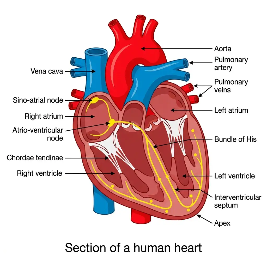
Anatomy of the Human Heart: A muscular four-chambered organ featuring right/left atria and ventricles, connected by a specialized conduction system.
🔬 Detailed Anatomical Description
The human heart is protected by a double-walled pericardium. The right side handles deoxygenated blood via the vena cava and pulmonary artery, while the left side pumps oxygenated blood via the pulmonary veins and aorta. Features the Sino-atrial Node (SAN, pacemaker), Atrio-ventricular Node (AVN), Bundle of His, and Chordae tendinae anchoring the valves.
XIV. CONDUCTING SYSTEM OF HEART
A NODAL TISSUE
| FEATURE | DETAIL |
|---|---|
| Specialised cardiac musculature | Nodal tissue ⭐⭐ |
| Property | Autoexcitable — can generate action potentials without external stimulus ⭐⭐⭐ |
B COMPONENTS AND SEQUENCE
| STRUCTURE | LOCATION / ROLE |
|---|---|
| SAN (Sino-atrial node) | Right upper corner of right atrium ⭐⭐⭐ |
| AVN (Atrio-ventricular node) | Lower left corner of right atrium near AV septum ⭐⭐⭐ |
| AV bundle / Bundle of His | Continues from AVN through AV septum ⭐⭐⭐ |
| Right and Left bundle branches | On top of interventricular septum ⭐⭐ |
| Purkinje fibres | Minute fibres in ventricular musculature ⭐⭐⭐ |
C PACEMAKER
| FEATURE | DETAIL |
|---|---|
| SAN action potential generation | 70–75/min — maximum in heart ⭐⭐⭐ |
| Role | Initiates and maintains rhythmic contractile activity ⭐⭐⭐ |
| Therefore SAN is called | Pacemaker ⭐⭐⭐ |
| Normal heart rate | 70–75 beats/min (average 72/min) ⭐⭐⭐ |
NEET 2024, NEET 2026: Conduction pathway = SAN → AVN → AV bundle → bundle branches → Purkinje fibres. RE-NEET 2026: The number of action potentials generated by the sino-atrial node (SAN) in a healthy human is 70–75 per minute.
XV. CARDIAC CYCLE
A BASIC SEQUENCE
| PHASE | EVENTS |
|---|---|
| Joint diastole | All 4 chambers relaxed; tricuspid and bicuspid valves open; semilunar valves closed ⭐⭐⭐ |
| Atrial systole | Both atria contract simultaneously ⭐⭐⭐ |
| Ventricular systole | Ventricles contract; atria relax ⭐⭐⭐ |
| Ventricular diastole | Ventricles relax; semilunar valves close ⭐⭐⭐ |
B JOINT DIASTOLE
| FEATURE | DETAIL |
|---|---|
| Condition | All four chambers relaxed ⭐⭐⭐ |
| AV valves | Open ⭐⭐⭐ |
| Semilunar valves | Closed ⭐⭐⭐ |
| Blood flow | Pulmonary veins → left atrium; vena cava → right atrium; then freely into ventricles ⭐⭐⭐ |
NEET 2022, 2024, NEET 2026: During joint diastole, blood moves freely from atria into ventricles (AV valves are open and semilunar valves are closed).
C ATRIAL SYSTOLE
| FEATURE | DETAIL |
|---|---|
| Action | Simultaneous contraction of both atria ⭐⭐⭐ |
| Result | Increases ventricular filling by about 30% ⭐⭐⭐ |
D VENTRICULAR SYSTOLE
| FEATURE | DETAIL |
|---|---|
| Coincides with | Atrial relaxation ⭐⭐ |
| Effect | Ventricular pressure rises ⭐⭐⭐ |
| Causes closure of | Tricuspid and bicuspid valves ⭐⭐⭐ |
| Causes opening of | Semilunar valves ⭐⭐⭐ |
| Blood enters | Pulmonary artery and aorta ⭐⭐⭐ |
E VENTRICULAR DIASTOLE
| FEATURE | DETAIL |
|---|---|
| Ventricles relax | Yes ⭐⭐ |
| Pressure falls | Yes ⭐⭐⭐ |
| Semilunar valves | Close ⭐⭐⭐ |
| Then AV valves | Open again as atrial pressure exceeds ventricular pressure ⭐⭐ |
F IMPORTANT NUMBERS
| PARAMETER | VALUE |
|---|---|
| Duration of one cardiac cycle | 0.8 second ⭐⭐⭐ |
| Stroke volume | ~70 mL blood pumped by each ventricle per cycle ⭐⭐⭐ |
| Cardiac output | Stroke volume × heart rate ⭐⭐⭐ |
| Average cardiac output | ~5 litres/min in healthy individual ⭐⭐⭐ |
XVI. HEART SOUNDS
| HEART SOUND | CAUSE |
|---|---|
| First heart sound (Lub) | Closure of tricuspid and bicuspid valves ⭐⭐⭐ |
| Second heart sound (Dub) | Closure of semilunar valves ⭐⭐⭐ |
NEET 2013, 2024: Lub = AV valves close; Dub = semilunar valves close.
XVII. ELECTROCARDIOGRAM (ECG)
A DEFINITION
| FEATURE | DETAIL |
|---|---|
| ECG | Graphical representation of electrical activity of heart during cardiac cycle ⭐⭐⭐ |
| Machine used | Electrocardiograph ⭐⭐ |
| Standard ECG | Obtained using 3 electrical leads (one to each wrist and left ankle) ⭐ |
B WAVES AND THEIR SIGNIFICANCE
| WAVE / COMPLEX | SIGNIFICANCE |
|---|---|
| P-wave | Depolarisation of atria → leads to contraction of both atria ⭐⭐⭐ |
| QRS complex | Depolarisation of ventricles; marks beginning of ventricular systole ⭐⭐⭐ |
| T-wave | Repolarisation of ventricles; end of T-wave marks end of systole ⭐⭐⭐ |
| T-P gap | Heart muscles are electrically silent ⭐⭐ |
C CLINICAL FACTS
| FEATURE | DETAIL |
|---|---|
| Heart beat rate determined by | Counting number of QRS complexes in a given time ⭐⭐⭐ |
| Reduction in T-wave size | Indicates coronary ischemia ⭐⭐ |
| ECG importance | Any deviation from normal shape indicates abnormality/disease ⭐⭐⭐ |
NEET 2023, 2024, NEET 2026: Heartbeat rate determined by QRS count. The T-P gap in an ECG represents the electrically silent phase when heart muscles are not showing electrical activity.
Standard ECG Wave
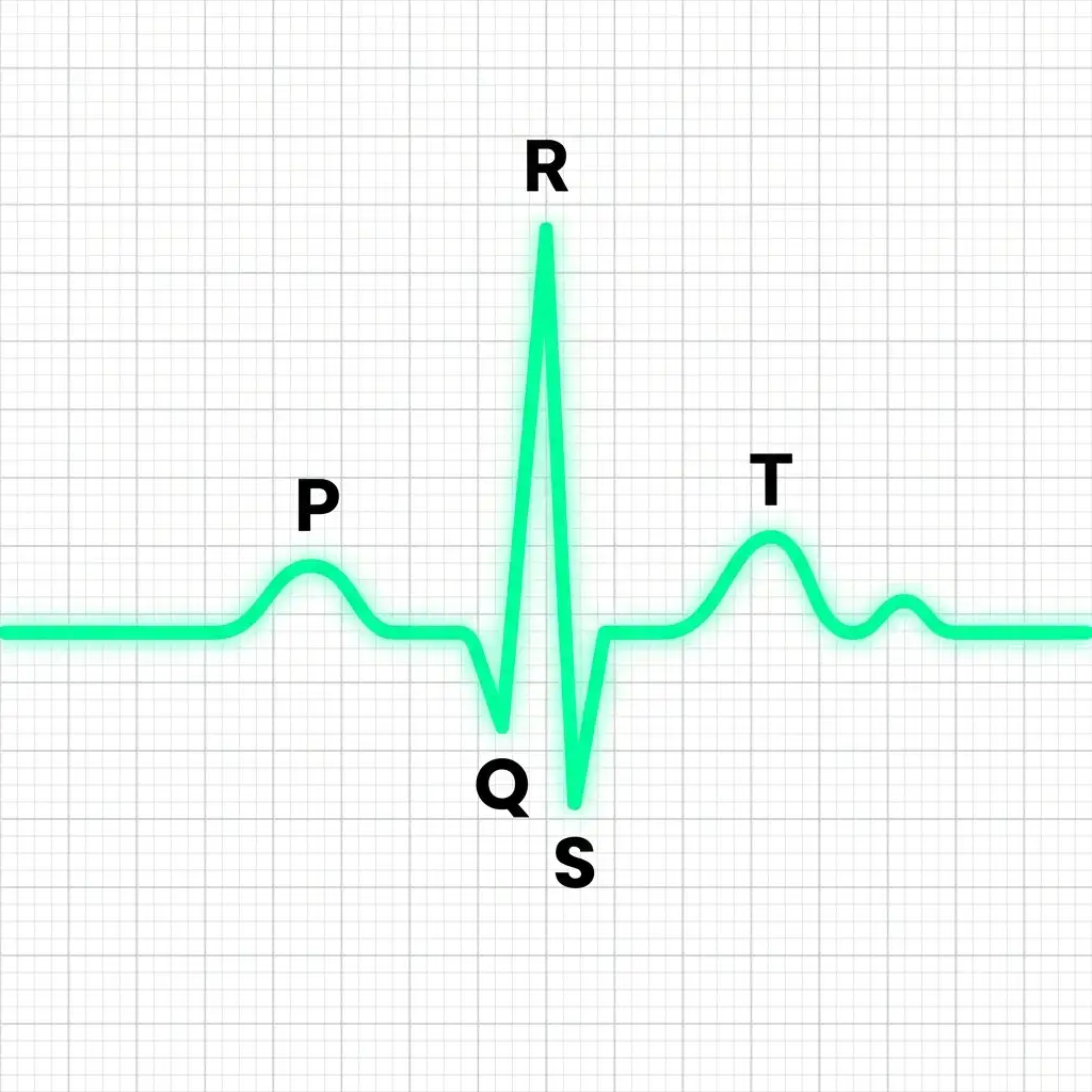
Standard ECG: A graphical representation of the electrical activity of the heart during a cardiac cycle, detailing waves P, Q, R, S, and T.
🔬 Detailed Anatomical Description
The ECG waves represent specific cardiac phases: the P-wave indicates atrial depolarization (contraction); the QRS complex represents ventricular depolarization (onset of ventricular contraction); and the T-wave represents ventricular repolarization (return to relaxed state). Vital diagnostic tool in clinical medicine.
XVIII. BLOOD VESSELS & DOUBLE CIRCULATION
A BLOOD VESSEL LAYERS
| LAYER | NAME | STRUCTURE |
|---|---|---|
| Inner | Tunica intima | Squamous endothelium ⭐⭐⭐ |
| Middle | Tunica media | Smooth muscle + elastic fibres ⭐⭐⭐ |
| Outer | Tunica externa | Fibrous connective tissue + collagen fibres ⭐⭐⭐ |
| KEY POINT | DETAIL |
|---|---|
| Tunica media in veins | Comparatively thin as compared to arteries ⭐⭐⭐ |
NEET 2013: Tunica media thinner in veins.
B DOUBLE CIRCULATION
| TYPE | PATHWAY |
|---|---|
| Pulmonary circulation | Right ventricle → pulmonary artery → lungs → pulmonary veins → left atrium ⭐⭐⭐ |
| Systemic circulation | Left ventricle → aorta → body tissues → venae cavae → right atrium ⭐⭐⭐ |
| SIGNIFICANCE | DETAIL |
|---|---|
| Double circulation | Keeps oxygenated and deoxygenated blood separate ⭐⭐⭐ |
| Present in | Birds, mammals, crocodiles ⭐⭐⭐ |
Schematic Plan of Blood Circulation
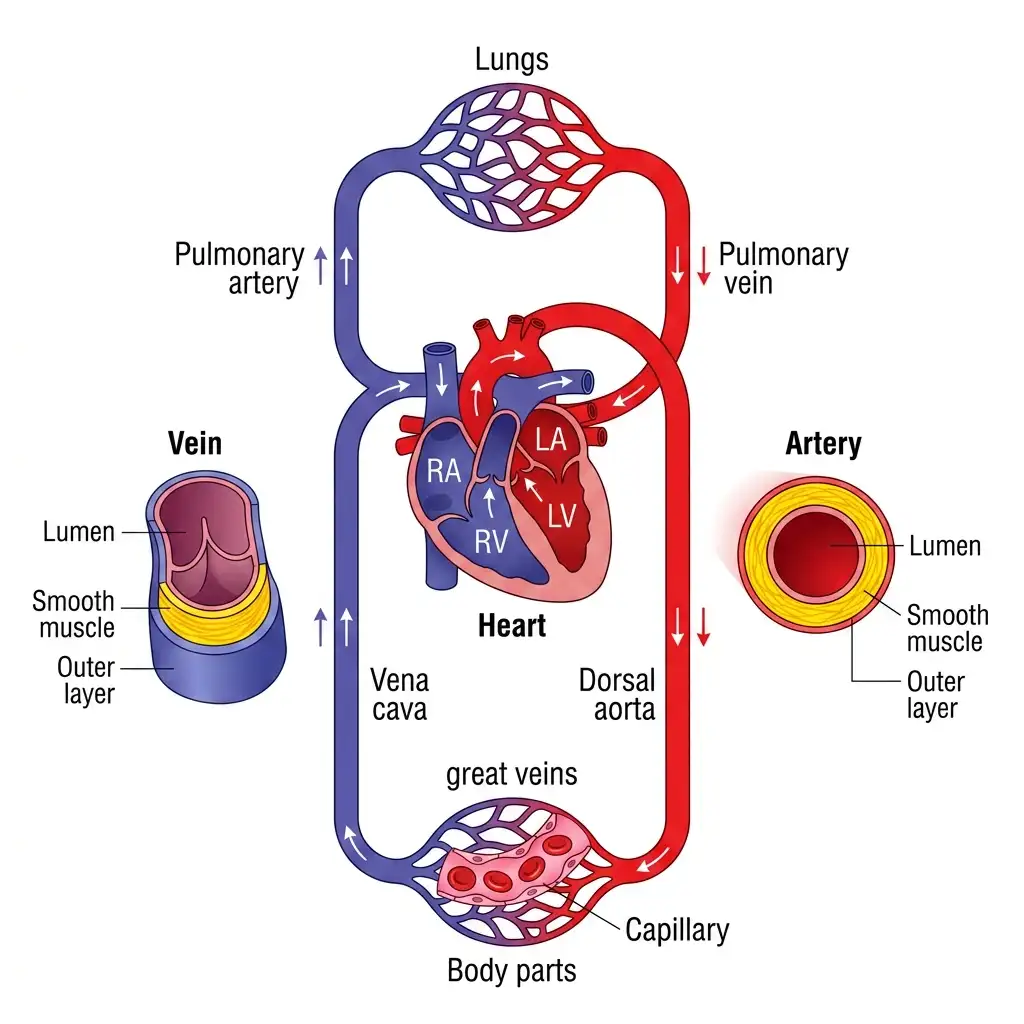
Double Circulation Schematic: Details the separation of oxygenated and deoxygenated blood through pulmonary and systemic circulation loops.
🔬 Detailed Anatomical Description
Double circulation involves two pathways: (1) Pulmonary Circulation: Deoxygenated blood is pumped from the right ventricle (RV) through the pulmonary artery to the lungs, and returns oxygenated to the left atrium (LA) via pulmonary veins. (2) Systemic Circulation: Oxygenated blood from the left ventricle (LV) goes via the aorta to body tissues and returns deoxygenated to the right atrium (RA) via vena cava. Artery walls are thicker with a narrow lumen; vein walls are thinner with a wider lumen.
C HEPATIC PORTAL SYSTEM
| FEATURE | DETAIL |
|---|---|
| Hepatic portal system | Unique vascular connection between digestive tract and liver ⭐⭐⭐ |
| Hepatic portal vein | Carries blood from intestine to liver before systemic circulation ⭐⭐⭐ |
NEET 2017, 2022: Hepatic portal system = intestine → liver.
D CORONARY CIRCULATION
| FEATURE | DETAIL |
|---|---|
| Coronary system | Special blood vessel system for circulation to and from cardiac musculature ⭐⭐ |
XIX. REGULATION OF CARDIAC ACTIVITY
| FEATURE | DETAIL |
|---|---|
| Heart is | Myogenic (auto-regulated by nodal tissue) ⭐⭐⭐ |
| Neural centre | Present in medulla oblongata ⭐⭐⭐ |
| Regulates via | Autonomic nervous system (ANS) ⭐⭐⭐ |
ANS Effects ⭐⭐⭐
| NERVE TYPE | EFFECT |
|---|---|
| Sympathetic nerves | Increase heart beat rate, strength of ventricular contraction, and cardiac output ⭐⭐⭐ |
| Parasympathetic nerves | Decrease heart beat rate, speed of conduction, and cardiac output ⭐⭐⭐ |
HORMONAL EFFECT
| FEATURE | DETAIL |
|---|---|
| Adrenal medullary hormones | Can increase cardiac output ⭐⭐ |
NEET 2020, NEET 2026: Parasympathetic signals strictly decrease heart rate and cardiac output. Sympathetic nerves increase heart rate, force of ventricular contraction and cardiac output.
XX. BLOOD PRESSURE
| FEATURE | DETAIL |
|---|---|
| Normal blood pressure | 120/80 mm Hg ⭐⭐⭐ |
| 120 mm Hg | Systolic pressure (pumping pressure) ⭐⭐⭐ |
| 80 mm Hg | Diastolic pressure (resting pressure) ⭐⭐⭐ |
| Hypertension | Repeated BP 140/90 mm Hg or higher ⭐⭐⭐ |
| Effect of high BP | Leads to heart diseases; affects brain and kidney ⭐⭐⭐ |
Additional BP Facts ⭐⭐
| FEATURE | DETAIL |
|---|---|
| BP in mammalian aorta maximum during | Systole of left ventricle ⭐⭐ |
| BP in pulmonary artery vs pulmonary vein | Pulmonary artery pressure > pulmonary vein pressure ⭐⭐ |
NEET 2026: Normal blood pressure is 120/80 mm Hg. Hypertension is repeatedly ≥140/90 mm Hg.
XXI. DISORDERS OF CIRCULATORY SYSTEM
A HYPERTENSION
| FEATURE | DETAIL |
|---|---|
| Definition | Blood pressure higher than normal (>140/90) ⭐⭐⭐ |
| Effects | Heart disease; affects brain and kidney ⭐⭐ |
B CORONARY ARTERY DISEASE (CAD) / ATHEROSCLEROSIS
| FEATURE | DETAIL |
|---|---|
| Other name | Atherosclerosis ⭐⭐⭐ |
| Affects | Vessels supplying blood to heart muscle ⭐⭐ |
| Cause | Deposition of calcium, fat, cholesterol and fibrous tissues ⭐⭐⭐ |
| Result | Narrowing of lumen of arteries ⭐⭐⭐ |
C ANGINA
| FEATURE | DETAIL |
|---|---|
| Other name | Angina pectoris ⭐⭐ |
| Symptom | Acute chest pain ⭐⭐⭐ |
| Cause | Not enough oxygen reaching heart muscle ⭐⭐⭐ |
| More common in | Middle-aged and elderly, but can occur at any age ⭐ |
NEET 2022, NEET 2026: Angina (angina pectoris) = acute chest pain that occurs when the heart muscle does not get enough oxygen.
D HEART FAILURE
| FEATURE | DETAIL |
|---|---|
| Definition | Heart not pumping blood effectively enough to meet body needs ⭐⭐⭐ |
| Also called | Congestive heart failure ⭐⭐ |
| Main symptom | Congestion of lungs ⭐ |
| Not same as | Cardiac arrest or heart attack ⭐⭐⭐ |
E CARDIAC ARREST
| FEATURE | DETAIL |
|---|---|
| Cardiac arrest | Heart stops beating ⭐⭐⭐ |
F HEART ATTACK
| FEATURE | DETAIL |
|---|---|
| Heart attack | Heart muscle suddenly damaged due to inadequate blood supply ⭐⭐⭐ |
XXII. RAPID REVISION — KEY COMPARISON TABLES
TABLE 1: Blood Components ⭐⭐⭐
| COMPONENT | DETAIL |
|---|---|
| Plasma | 55% of blood; 90–92% water; 6–8% proteins |
| Formed Elements | RBCs, WBCs, Platelets |
TABLE 2: Plasma Proteins ⭐⭐⭐
| PROTEIN | FUNCTION |
|---|---|
| Fibrinogen | Clotting / coagulation |
| Globulins | Defense / antibodies |
| Albumins | Osmotic balance |
TABLE 3: Formed Elements ⭐⭐⭐
| ELEMENT | COUNT | FUNCTION |
|---|---|---|
| RBCs | 5–5.5 million/mm³ | Respiratory gas transport |
| WBCs | 6000–8000/mm³ | Defense / immune response |
| Platelets | 1.5–3.5 lakh/mm³ | Clotting |
TABLE 4: WBC Types and Percentages ⭐⭐⭐
| TYPE | CATEGORY | % | FUNCTION |
|---|---|---|---|
| Neutrophils | Granulocyte | 60–65 | Phagocytosis |
| Eosinophils | Granulocyte | 2–3 | Resist infection; allergy |
| Basophils | Granulocyte | 0.5–1 | Histamine, serotonin, heparin; inflammation |
| Lymphocytes | Agranulocyte | 20–25 | Immune response |
| Monocytes | Agranulocyte | 6–8 | Phagocytosis |
TABLE 5: ABO Blood Groups ⭐⭐⭐
| GROUP | ANTIGEN ON RBC | ANTIBODY IN PLASMA | CAN RECEIVE FROM |
|---|---|---|---|
| A | A | Anti-B | A, O |
| B | B | Anti-A | B, O |
| AB | A, B | Nil | A, B, AB, O |
| O | Nil | Anti-A, Anti-B | O |
TABLE 6: Open vs Closed Circulation ⭐⭐⭐
| FEATURE | OPEN | CLOSED |
|---|---|---|
| Organisms | Arthropods, Molluscs | Annelids, Chordates |
| Fluid pathway | Into sinuses / body cavity | Closed vessels |
| Regulation | Less precise | More precise |
TABLE 7: Vertebrate Hearts ⭐⭐⭐
| GROUP | CHAMBERS | CIRCULATION |
|---|---|---|
| Fish | 2 | Single |
| Amphibians | 3 | Incomplete double |
| Reptiles (except crocodile) | 3 | Incomplete double |
| Crocodiles, Birds, Mammals | 4 | Complete double |
TABLE 8: Heart Valves ⭐⭐⭐
| VALVE | LOCATION |
|---|---|
| Tricuspid | Right atrium ↔ Right ventricle |
| Bicuspid / Mitral | Left atrium ↔ Left ventricle |
| Semilunar | Ventricles ↔ Pulmonary artery / Aorta |
TABLE 9: Conducting System ⭐⭐⭐
| STRUCTURE | SEQUENCE |
|---|---|
| SAN | 1st |
| AVN | 2nd |
| AV bundle | 3rd |
| Bundle branches | 4th |
| Purkinje fibres | 5th |
TABLE 10: Cardiac Cycle Facts ⭐⭐⭐
| PARAMETER | VALUE |
|---|---|
| Duration of cardiac cycle | 0.8 sec |
| Stroke volume | ~70 mL |
| Heart rate | ~72/min |
| Cardiac output | ~5 L/min |
TABLE 11: ECG Waves ⭐⭐⭐
| WAVE | MEANING |
|---|---|
| P-wave | Atrial depolarisation |
| QRS complex | Ventricular depolarisation |
| T-wave | Ventricular repolarisation |
| T-P gap | Electrically silent phase |
TABLE 12: Double Circulation ⭐⭐⭐
| TYPE | ROUTE |
|---|---|
| Pulmonary | Right ventricle → lungs → left atrium |
| Systemic | Left ventricle → body → right atrium |
TABLE 13: Disorders ⭐⭐⭐
| DISORDER | MEANING |
|---|---|
| Hypertension | BP > 140/90 mm Hg |
| CAD / Atherosclerosis | Narrowing of coronary arteries due to deposits |
| Angina | Chest pain due to low O₂ to heart muscle |
| Heart failure | Heart not pumping effectively |
| Cardiac arrest | Heart stops beating |
| Heart attack | Sudden damage to heart muscle due to poor blood supply |
XXIII. KEY NUMBERS — QUICK REFERENCE
| PARAMETER | VALUE |
|---|---|
| Plasma percentage | 55% |
| Plasma water | 90–92% |
| Plasma proteins | 6–8% |
| RBC count | 5–5.5 million/mm³ |
| WBC count | 6000–8000/mm³ |
| Platelet count | 1,50,000–3,50,000/mm³ |
| Haemoglobin | 12–16 g/100 mL blood |
| RBC lifespan | 120 days |
| SAN rate | 70–75/min |
| Normal heart rate | 72/min |
| Cardiac cycle duration | 0.8 sec |
| Stroke volume | 70 mL |
| Cardiac output | 5 L/min |
| Normal BP | 120/80 mm Hg |
| Hypertension | 140/90 mm Hg or above |
| Neutrophils | 60–65% |
| Lymphocytes | 20–25% |
| Monocytes | 6–8% |
| Eosinophils | 2–3% |
| Basophils | 0.5–1% |
XXIV. COMMON EXAM TRAPS — QUICK REFERENCE
| TRAP / QUESTION | CORRECT ANSWER |
|---|---|
| Blood is what type of tissue? | Special connective tissue |
| Plasma without clotting factors? | Serum |
| Most abundant blood cell? | RBC |
| RBC formed in adults where? | Red bone marrow |
| RBC graveyard? | Spleen |
| Adult mammalian RBC nucleus? | Absent |
| Least abundant WBC? | Basophils |
| Most abundant WBC? | Neutrophils |
| Phagocytic WBCs? | Neutrophils and Monocytes |
| Immune response WBC? | Lymphocytes |
| Basophils secrete? | Histamine, serotonin, heparin |
| Platelets are complete cells? | No, cell fragments |
| Platelets produced from? | Megakaryocytes |
| Universal donor? | O group |
| Universal recipient? | AB group / AB⁺ |
| AB blood has which antibodies? | Nil |
| O blood has which antigens? | Nil |
| Rh incompatibility occurs in? | Rh⁻ mother, Rh⁺ foetus |
| First pregnancy in Rh incompatibility? | Usually normal |
| Foetal disease due to Rh incompatibility? | Erythroblastosis foetalis |
| Enzyme converting fibrinogen to fibrin? | Thrombin |
| Clot made of thrombin threads? | No, fibrin threads |
| Ca²⁺ role? | Important in clotting |
| Lymph colour? | Colourless |
| Fats absorbed through? | Lacteals of intestinal villi |
| Open circulation found in? | Arthropods, Molluscs |
| Closed circulation found in? | Annelids, Chordates |
| Fish circulation? | Single circulation |
| Amphibian / reptile heart? | 3-chambered |
| Mammal / bird / crocodile heart? | 4-chambered |
| Heart origin? | Mesodermal |
| Pacemaker of heart? | SAN |
| Why SAN pacemaker? | Maximum AP generation (70–75/min) |
| Tricuspid location? | RA–RV |
| Mitral / Bicuspid location? | LA–LV |
| Semilunar valves location? | Ventricles to pulmonary artery / aorta |
| During joint diastole AV valves? | Open |
| During joint diastole semilunar valves? | Closed |
| Lub sound due to? | Closure of tricuspid + bicuspid |
| Dub sound due to? | Closure of semilunar |
| P-wave? | Atrial depolarisation |
| QRS? | Ventricular depolarisation |
| T-wave? | Ventricular repolarisation |
| Heart rate from ECG determined by? | QRS count |
| Pulmonary circulation starts from? | Right ventricle |
| Systemic circulation starts from? | Left ventricle |
| Hepatic portal vein carries blood from? | Intestine to liver |
| Heart is neurogenic or myogenic? | Myogenic |
| Sympathetic effect? | Increases heart rate and cardiac output |
| Parasympathetic effect? | Decreases heart rate and cardiac output |
| Normal BP? | 120/80 mm Hg |
| Hypertension? | 140/90 or higher |
| CAD also called? | Atherosclerosis |
| Angina means? | Acute chest pain due to less O₂ to heart muscle |
| Heart failure same as cardiac arrest? | No |
| Cardiac arrest? | Heart stops beating |
| Heart attack? | Sudden damage due to inadequate blood supply |
- RE-NEET 2026: SAN generates 70–75 action potentials/min.
- NEET 2026: Conducting pathway = SAN → AVN → AV bundle → bundle branches → Purkinje fibres.
- NEET 2026: During joint diastole, blood flows freely from atria into ventricles (AV valves open, semilunar closed).
- NEET 2026 context / reinforced: Rh incompatibility = Rh⁻ mother, Rh⁺ foetus; erythroblastosis foetalis in 2nd pregnancy.
- NEET 2026 / reinforced: Blood clot formed by fibrin threads (not thrombin threads).
- NEET 2026: Heart rate determined by counting QRS complexes on ECG; T-P gap = electrically silent phase.
- NEET 2026: Sympathetic nerves increase heart rate, contraction strength & cardiac output; parasympathetic decrease heart rate & cardiac output.
- NEET 2026: Normal BP = 120/80 mm Hg; Hypertension = ≥140/90 mm Hg; Angina = acute chest pain due to low O₂ to heart muscle.