I. RESPIRATORY ORGANS
A RESPIRATORY ORGANS IN DIFFERENT ORGANISMS
Invertebrates ⭐⭐
| ORGANISM | RESPIRATORY ORGAN / MECHANISM |
|---|---|
| Sponges, Coelenterates, Flatworms | Exchange O₂ & CO₂ by simple diffusion over entire body surface ⭐ |
| Earthworms | Through moist cuticle (Cutaneous respiration) ⭐ |
| Insects | Network of tracheal tubes (transport atmospheric air within body) ⭐ |
| Aquatic Arthropods & Molluscs | Gills (Branchial respiration) ⭐ |
| Terrestrial forms | Lungs (Pulmonary respiration) ⭐ |
Vertebrates ⭐⭐
| ORGANISM | RESPIRATORY ORGAN |
|---|---|
| Fishes | Gills ⭐ |
| Amphibians, Reptiles, Birds, Mammals | Lungs ⭐ |
| Amphibians (e.g., Frog) | ALSO through moist skin (Cutaneous respiration) ⭐⭐ |
| KEY FACT | DETAIL |
|---|---|
| Mechanism of breathing depends on | Habitat and Level of organisation ⭐ |
B HUMAN RESPIRATORY SYSTEM — PATHWAY
C KEY STRUCTURES
| STRUCTURE | DETAIL |
|---|---|
| External nostrils | Open above the upper lips ⭐ |
| Nasal chamber | Opens into pharynx ⭐ |
| Pharynx | Common passage for food and air ⭐ |
| Larynx | Cartilaginous box; helps in sound production = "Sound box" ⭐ |
| Epiglottis | Thin elastic cartilaginous flap → covers glottis during swallowing → prevents entry of food into larynx ⭐⭐ |
| Trachea | Straight tube extending up to mid-thoracic cavity ⭐ |
| Trachea divides at | Level of 5th thoracic vertebra into right & left primary bronchi ⭐⭐ |
| Cartilaginous rings | Trachea, primary, secondary, tertiary bronchi and initial bronchioles are supported by incomplete cartilaginous rings ⭐ |
| Terminal bronchioles | Give rise to alveoli ⭐ |
| Alveoli | Very thin, irregular-walled, vascularised, bag-like structures ⭐ |
| Lungs | Branching network of bronchi, bronchioles and alveoli ⭐ |
D LUNGS & PLEURA
| FEATURE | DETAIL |
|---|---|
| Number of lungs | Two (pair) ⭐ |
| Covering | Double-layered pleura ⭐ |
| Pleural fluid | Between the two layers → reduces friction on lung surface ⭐ |
| Outer pleural membrane | In close contact with thoracic lining ⭐ |
| Inner pleural membrane | In contact with lung surface ⭐ |
| Lung collapse prevention | Negative intrapleural pressure pulling at lung walls prevents collapse between breaths ⭐ |
NEET 2016, NEET 2026 context / reinforced: Lungs don't collapse between breaths because of negative intrapleural pressure pulling at lung walls.
Human Respiratory System
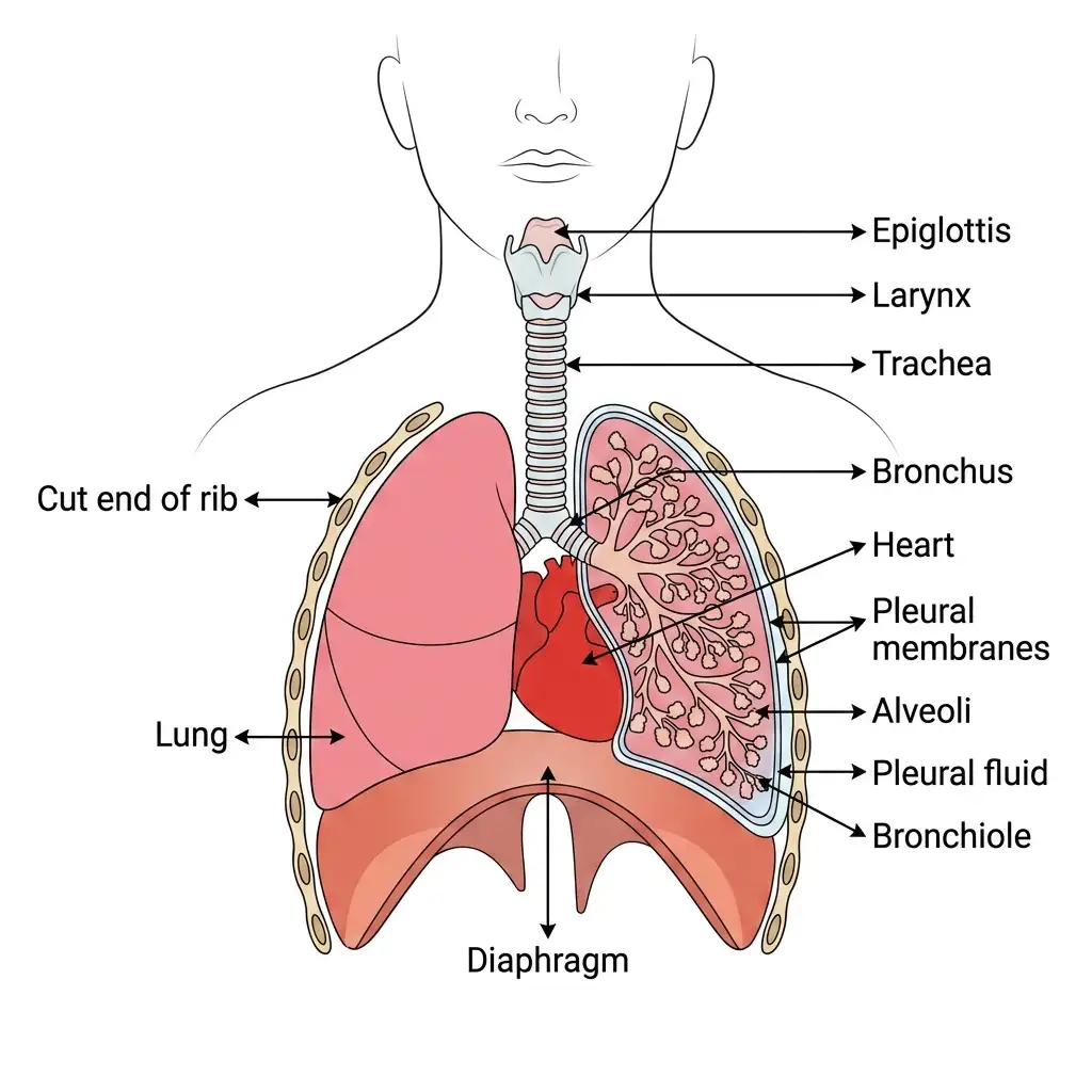
Human Respiratory System: A sectional view showing epiglottis, larynx, trachea, lungs with branching bronchus/bronchioles, and diaphragm.
🔬 Detailed Anatomical Description
The human respiratory tract begins at the nose, moving down to the pharynx, larynx (sound box), and trachea. The trachea divides into primary bronchi entering the left and right lungs. Inside each lung, bronchi branch into bronchioles terminating in tiny vascularized sacs called alveoli. Lungs are protected by a double-layered pleural membrane with friction-reducing pleural fluid, supported at the base by the skeletal diaphragm muscle.
E CONDUCTING PART vs RESPIRATORY PART
| PART | EXTENT | FUNCTION |
|---|---|---|
| Conducting Part | External nostrils → Terminal bronchioles ⭐⭐ | (1) Transports air to alveoli; (2) Clears foreign particles; (3) Humidifies air; (4) Brings air to body temperature ⭐⭐ |
| Respiratory / Exchange Part | Alveoli and their ducts ⭐⭐ | Actual site of diffusion of O₂ and CO₂ between blood and atmospheric air ⭐⭐ |
NEET 2013, 2022, NEET 2026 context: Conducting part = nostrils to terminal bronchioles; functions — transport, clear, humidify, body temperature. Exchange part = alveoli; actual diffusion site.
F THORACIC CHAMBER
| BOUNDARY | FORMED BY |
|---|---|
| Dorsally | Vertebral column ⭐ |
| Ventrally | Sternum ⭐ |
| Laterally | Ribs ⭐ |
| Lower side | Diaphragm (dome-shaped) ⭐ |
| KEY PRINCIPLE | DETAIL |
|---|---|
| Any change in thoracic cavity volume | Reflected in lung (pulmonary) cavity volume ⭐ |
| We cannot | Directly alter pulmonary volume ⭐ |
| Thoracic chamber is | Anatomically air-tight ⭐ |
II. MECHANISM OF BREATHING
A STEPS OF RESPIRATION
| STEP | DETAIL |
|---|---|
| 1 | Breathing / Pulmonary ventilation (air drawn in & CO₂-rich air released out) ⭐ |
| 2 | Diffusion of gases (O₂ & CO₂) across alveolar membrane ⭐ |
| 3 | Transport of gases by blood ⭐ |
| 4 | Diffusion of O₂ & CO₂ between blood and tissues ⭐ |
| 5 | Utilisation of O₂ by cells for catabolic reactions → release of CO₂ (cellular respiration) ⭐ |
NEET 2023: Know the complete sequence of respiration (5 steps).
B INSPIRATION & EXPIRATION
| FEATURE | INSPIRATION | EXPIRATION |
|---|---|---|
| Definition | Atmospheric air drawn IN ⭐ | Alveolar air released OUT ⭐ |
| Pressure condition | Intra-pulmonary pressure < Atmospheric pressure ⭐⭐ | Intra-pulmonary pressure > Atmospheric pressure ⭐⭐ |
| Diaphragm | Contracts (flattens) ⭐ | Relaxes (returns to dome shape) ⭐ |
| External intercostal muscles | Contract → lift ribs & sternum ⭐ | Relax → ribs & sternum return to normal ⭐ |
| Thoracic volume | Increases ⭐ | Decreases ⭐ |
| Pulmonary volume | Increases ⭐ | Decreases ⭐ |
| Intra-pulmonary pressure | Decreases (below atmospheric) ⭐ | Increases (above atmospheric) ⭐ |
NEET 2019, 2020, NEET 2026 context: Inspiration — contraction of diaphragm (flattens) + external intercostal muscles (lift ribs & sternum) → thoracic volume increases in antero-posterior and dorso-ventral axes → intra-pulmonary pressure decreases → air flows in. Expiration: relaxation of diaphragm (dome shape) + external intercostal muscles → thoracic volume decreases → intra-pulmonary pressure increases → air flows out.
Axis of Volume Change During Inspiration ⭐⭐
| ACTION | AXIS |
|---|---|
| Contraction of diaphragm | Increases thoracic volume in antero-posterior axis ⭐ |
| Contraction of external intercostal muscles | Increases thoracic volume in dorso-ventral axis ⭐ |
| Overall | Both axes → overall thoracic volume increases ⭐ |
Additional Facts ⭐
| FEATURE | DETAIL |
|---|---|
| Normal breathing rate | 12–16 times/minute (healthy human) ⭐ |
| Additional muscles | Abdominal muscles can increase strength of inspiration & expiration ⭐ |
| Volume estimation | Using a spirometer (clinical assessment of pulmonary functions) ⭐ |
Mechanism of Breathing
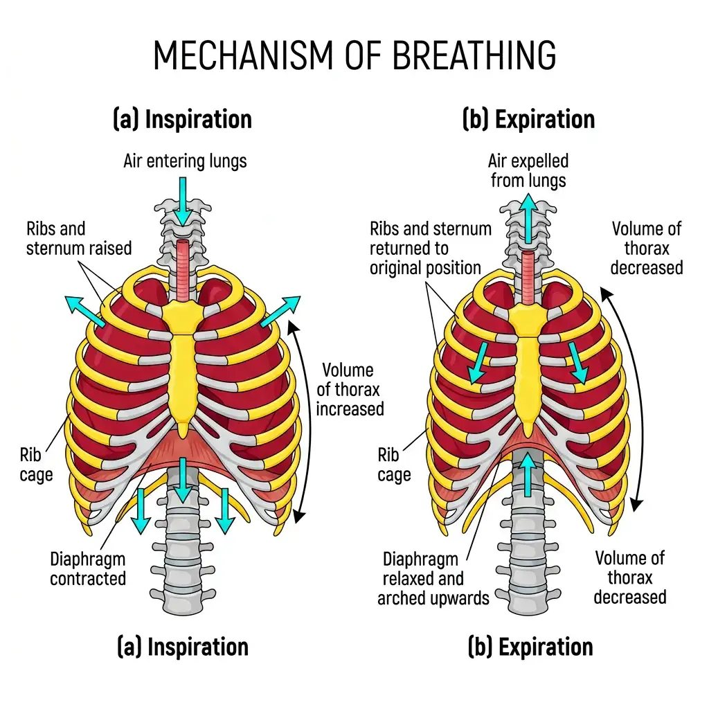
Mechanism of Breathing: (a) Inspiration is an active process driven by diaphragm contraction, and (b) Expiration is a passive relaxation phase.
🔬 Detailed Anatomical Description
Breathing relies on pressure gradients between the lungs and atmosphere. (a) Inspiration: The diaphragm contracts and flattens, while external intercostal muscles lift the ribs and sternum. This expands thoracic volume, drops intra-pulmonary pressure, and draws air inside. (b) Expiration: Muscles relax, the diaphragm arches upwards, thoracic volume shrinks, increasing pressure and pushing air out of the lungs.
III. RESPIRATORY VOLUMES & CAPACITIES
A RESPIRATORY VOLUMES
| VOLUME | ABBR. | DEFINITION | VALUE |
|---|---|---|---|
| Tidal Volume | TV | Volume of air inspired/expired during normal respiration ⭐ | ~500 mL ⭐⭐ |
| Inspiratory Reserve Volume | IRV | Additional volume inspired by forcible inspiration ⭐ | 2500–3000 mL ⭐ |
| Expiratory Reserve Volume | ERV | Additional volume expired by forcible expiration ⭐ | 1000–1100 mL ⭐ |
| Residual Volume | RV | Volume remaining in lungs even after forcible expiration ⭐⭐ | 1100–1200 mL ⭐⭐ |
| ADDITIONAL FACT | DETAIL |
|---|---|
| Air per minute (normal) | TV × breathing rate = 500 mL × 12–16 = 6000–8000 mL/minute ⭐ |
| Residual volume significance | Prevents alveoli from collapsing ⭐ |
NEET 2016, 2017, 2018, 2024, NEET 2026 context: TV ≈ 500 mL; IRV = 2500–3000 mL; ERV = 1000–1100 mL; RV = 1100–1200 mL (air remaining after forcible expiration).
B PULMONARY CAPACITIES
| CAPACITY | ABBR. | FORMULA | DETAIL |
|---|---|---|---|
| Inspiratory Capacity | IC | TV + IRV ⭐ | Total volume a person can inspire after normal expiration |
| Expiratory Capacity | EC | TV + ERV ⭐⭐ | Total volume a person can expire after normal inspiration |
| Functional Residual Capacity | FRC | ERV + RV ⭐ | Volume remaining after normal expiration |
| Vital Capacity | VC | ERV + TV + IRV ⭐⭐⭐ | Maximum volume a person can breathe in after forced expiration (or out after forced inspiration) |
| Total Lung Capacity | TLC | RV + ERV + TV + IRV = RV + VC ⭐⭐ | Total volume at end of forced inspiration |
NEET 2019, 2020, 2022, 2023, 2024, NEET 2026: EC = TV + ERV; VC = ERV + TV + IRV; TLC = RV + ERV + TV + IRV = RV + VC; IC = TV + IRV; FRC = ERV + RV. Vital Capacity definition — maximum volume after forced expiration.
IV. EXCHANGE OF GASES
A PRIMARY SITE
| FEATURE | DETAIL |
|---|---|
| Primary site of gas exchange | Alveoli ⭐⭐ |
| Exchange also occurs between | Blood and tissues ⭐ |
| Mechanism | Simple diffusion ⭐ |
B FACTORS AFFECTING DIFFUSION
| FACTOR | DETAIL |
|---|---|
| 1. Pressure / Concentration gradient | Main driving force ⭐ |
| 2. Solubility of gases | CO₂ solubility is 20–25 times higher than O₂ → diffuses faster ⭐⭐ |
| 3. Thickness of membrane | Diffusion membrane thickness much less than 1 mm ⭐ |
C PARTIAL PRESSURES (mm Hg)
| GAS | ATMOSPHERIC AIR | ALVEOLI | BLOOD (DEOXY) | BLOOD (OXY) | TISSUES |
|---|---|---|---|---|---|
| O₂ | 159 | 104 | 40 | 95 | 40 |
| CO₂ | 0.3 | 40 | 45 | 40 | 45 |
Gradient Direction ⭐⭐
| GAS | GRADIENT |
|---|---|
| O₂ | Alveoli (104) → Deoxygenated blood (40) → Tissues (40) — favours O₂ flow from alveoli to tissues ⭐ |
| CO₂ | Tissues (45) → Blood (45) → Alveoli (40) — favours CO₂ flow from tissues to alveoli ⭐ |
NEET 2016, 2021, NEET 2026 context: Know all partial pressure values; pO₂ in alveoli (104) > deoxygenated blood (40). Partial pressures (mm Hg): pO₂ alveoli = 104; deoxygenated blood = 40; tissues = 40; pCO₂ alveoli = 40; tissues = 45.
Exchange and Transport of Gases
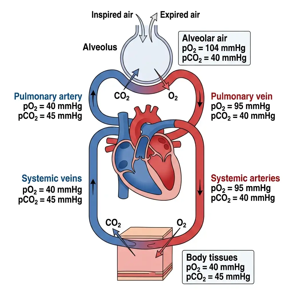
Gas Exchange & Transport: Depicts the partial pressures (pO2 & pCO2) regulating passive diffusion of respiratory gases across membranes.
🔬 Detailed Anatomical Description
Gas exchange occurs by simple passive diffusion governed by partial pressure gradients. In the alveolus, pO2 is high (104 mmHg) and pCO2 is low (40 mmHg), causing O2 to diffuse into the blood and CO2 to diffuse out. At body tissues, respiration consumes O2 (pO2=40) and produces CO2 (pCO2=45), reversing the gradient so O2 enters tissue cells and CO2 enters the bloodstream.
D DIFFUSION MEMBRANE (3 LAYERS)
| LAYER | DETAIL |
|---|---|
| 1 | Thin squamous epithelium of alveoli ⭐ |
| 2 | Endothelium of alveolar capillaries ⭐ |
| 3 | Basement substance (thin basement membrane supporting squamous epithelium + basement membrane surrounding capillary endothelial cells) ⭐ |
| KEY FACT | DETAIL |
|---|---|
| Total thickness | Much less than 1 mm (< 1 millimetre) ⭐ |
| All factors in body | Favourable for diffusion of O₂ from alveoli → tissues and CO₂ from tissues → alveoli ⭐ |
NEET 2016, NEET 2026 context: Primary site of gas exchange = alveoli. Diffusion membrane has 3 layers and thickness much less than 1 mm. CO₂ is 20–25 times more soluble than O₂.
Section of Alveolus & Capillary
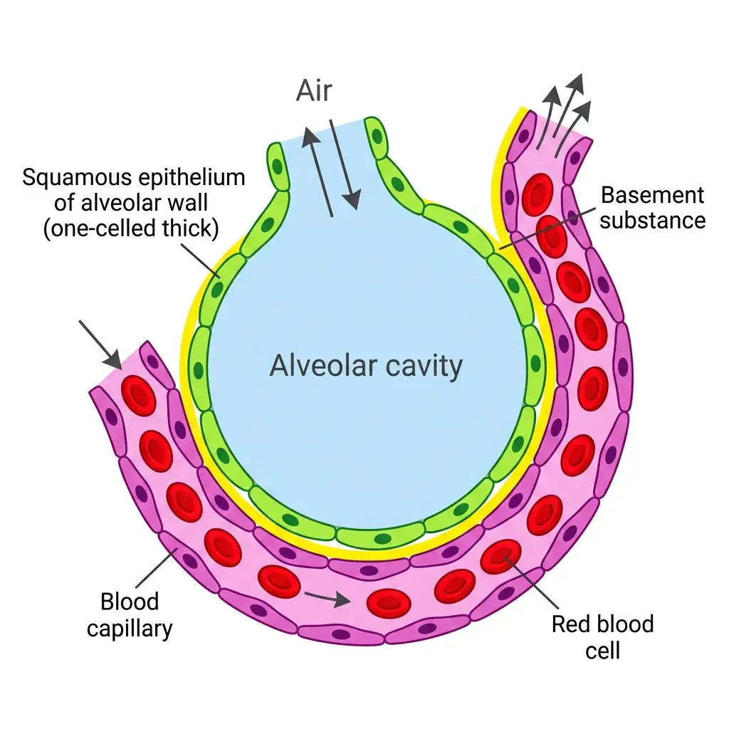
The Diffusion Membrane: Composed of three thin layers: squamous epithelium of alveolus, basement substance, and capillary endothelium.
🔬 Detailed Anatomical Description
The respiratory diffusion membrane is highly specialized for gas transfer, with a total thickness of less than a millimeter. It is composed of: (1) Single-layered thin squamous epithelium of the alveolar wall; (2) An acellular non-living basement substance; and (3) Single-layered endothelium of the pulmonary blood capillary. Red blood cells flow through the capillary in close proximity, enabling rapid oxygenation.
V. TRANSPORT OF GASES
A OVERVIEW OF GAS TRANSPORT
Oxygen Transport ⭐⭐⭐
| MODE | PERCENTAGE |
|---|---|
| By RBCs (as oxyhaemoglobin) | ~97% ⭐⭐ |
| Dissolved in plasma | ~3% ⭐ |
Carbon Dioxide Transport ⭐⭐⭐
| MODE | PERCENTAGE |
|---|---|
| As Bicarbonate (HCO₃⁻) | ~70% ⭐⭐⭐ |
| By RBCs (as carbamino-haemoglobin) | ~20–25% ⭐⭐ |
| Dissolved in plasma | ~7% ⭐ |
NEET 2014, 2024, NEET 2026 context: CO₂ transport — ~70% as bicarbonate; ~20-25% as carbamino-haemoglobin; ~7% dissolved in plasma. O₂ transport — ~97% as oxyhaemoglobin, ~3% dissolved in plasma.
B TRANSPORT OF OXYGEN — DETAILS
| FEATURE | DETAIL |
|---|---|
| Haemoglobin | Red-coloured, iron-containing pigment present in RBCs ⭐ |
| Binding | O₂ binds Hb in a reversible manner → Oxyhaemoglobin ⭐ |
| Capacity | Each Hb molecule can carry maximum 4 molecules of O₂ ⭐ |
| Primary factor | Binding primarily depends on partial pressure of O₂ (pO₂) ⭐⭐ |
| Other factors | pCO₂, H⁺ concentration, Temperature ⭐ |
C OXYGEN DISSOCIATION CURVE
| FEATURE | DETAIL |
|---|---|
| Definition | Curve obtained when % saturation of Hb with O₂ is plotted against pO₂ ⭐⭐ |
| Shape | Sigmoid (S-shaped) curve ⭐⭐ |
Oxygen Dissociation Curve
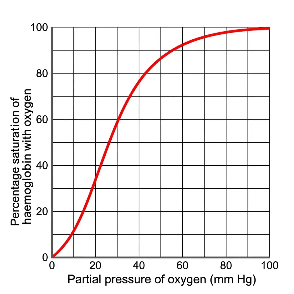
Oxygen-Haemoglobin Dissociation Curve: S-shaped (sigmoid) curve representing oxygen-hemoglobin binding dynamics under physiological conditions.
🔬 Detailed Anatomical Description
Under normal physiological conditions, oxygen binds reversibly to hemoglobin in a cooperative manner, generating a sigmoid curve. At the lungs, high pO2, low pCO2, low H+ concentration, and lower temperature facilitate oxyhemoglobin formation. In tissues, low pO2, high pCO2, high H+ (acidity), and high temperature encourage oxygen dissociation, releasing O2 for metabolic activity.
D CONDITIONS IN ALVEOLI vs TISSUES
| FACTOR | IN ALVEOLI | IN TISSUES |
|---|---|---|
| pO₂ | HIGH ⭐ | LOW ⭐ |
| pCO₂ | LOW ⭐ | HIGH ⭐ |
| H⁺ concentration | LOW (higher pH) ⭐ | HIGH (lower pH) ⭐ |
| Temperature | LOWER ⭐ | HIGHER ⭐ |
| Favours | Formation of oxyhaemoglobin ⭐⭐ | Dissociation of oxygen (from oxyhaemoglobin) ⭐⭐ |
NEET 2020, 2021, 2024: Alveoli — high pO₂, low pCO₂, low H⁺, low temp → oxyhaemoglobin formation. Tissues — low pO₂, high pCO₂, high H⁺, high temp → O₂ dissociation.
Bohr Effect ⭐⭐
| FEATURE | DETAIL |
|---|---|
| Definition | Reduction in blood pH (increase in H⁺) decreases affinity of Hb for O₂ ⭐ |
| Curve shift | Shifts O₂ dissociation curve to the right ⭐ |
NEET 2016, NEET 2026 context: Bohr Effect — ↑H⁺ (lower pH) → ↓ Hb-O₂ affinity → curve shifts right.
E O₂ DELIVERY TO TISSUES
| FEATURE | DETAIL |
|---|---|
| Every 100 mL of oxygenated blood | Delivers around 5 mL of O₂ to tissues ⭐⭐ |
| Under | Normal physiological conditions ⭐ |
NEET 2022, NEET 2026 context: Every 100 mL of oxygenated blood delivers ~5 mL of O₂ to tissues. Every 100 mL of deoxygenated blood delivers ~4 mL of CO₂ to alveoli.
F TRANSPORT OF CARBON DIOXIDE — DETAILS
| FEATURE | DETAIL |
|---|---|
| CO₂ carried by Hb as | Carbamino-haemoglobin (~20–25%) ⭐ |
| Binding related to | Partial pressure of CO₂ (pCO₂) ⭐ |
| Major factor affecting binding | pO₂ ⭐ |
CO₂ Binding at Different Sites ⭐⭐
| SITE | CONDITIONS | WHAT HAPPENS |
|---|---|---|
| Tissues | High pCO₂ + Low pO₂ | More binding of CO₂ to Hb (carbamino-Hb formation) ⭐ |
| Alveoli | Low pCO₂ + High pO₂ | Dissociation of CO₂ from carbamino-Hb → CO₂ released ⭐ |
G BICARBONATE TRANSPORT (70%) — CARBONIC ANHYDRASE
| FEATURE | DETAIL |
|---|---|
| Enzyme | Carbonic anhydrase ⭐⭐ |
| Location | Very high concentration in RBCs; minute quantities in plasma ⭐ |
| Reaction | CO₂ + H₂O ⇌ H₂CO₃ ⇌ HCO₃⁻ + H⁺ ⭐⭐ |
| Bidirectional | Enzyme facilitates reaction in BOTH directions ⭐ |
| AT TISSUES | AT ALVEOLI |
|---|---|
| pCO₂ HIGH (due to catabolism) | pCO₂ LOW |
| CO₂ diffuses into blood → forms HCO₃⁻ (bicarbonate) + H⁺ ⭐ | Reaction proceeds in opposite direction → forms CO₂ + H₂O ⭐ |
| CO₂ trapped as bicarbonate ⭐ | CO₂ released out from alveoli ⭐ |
NEET 2020, NEET 2026 context: Carbonic anhydrase is present in very high concentration in RBCs (minute quantities in plasma). It facilitates CO₂ + H₂O ⇌ H₂CO₃ ⇌ HCO₃⁻ + H⁺ in both directions.
H CO₂ DELIVERY TO ALVEOLI
| FEATURE | DETAIL |
|---|---|
| Every 100 mL of deoxygenated blood | Delivers approximately 4 mL of CO₂ to alveoli ⭐ |
VI. REGULATION OF RESPIRATION
A NEURAL REGULATION
| CENTRE | LOCATION | FUNCTION |
|---|---|---|
| Respiratory Rhythm Centre | Medulla region of brain ⭐⭐ | Primarily responsible for regulation of respiration ⭐ |
| Pneumotaxic Centre | Pons region of brain ⭐⭐ | Moderates function of respiratory rhythm centre; can reduce duration of inspiration → alters respiratory rate ⭐ |
| Chemo-sensitive area | Adjacent to rhythm centre (in medulla) ⭐ | Highly sensitive to CO₂ and H⁺ → activates rhythm centre for adjustments ⭐⭐ |
NEET 2020, NEET 2026 context: Pneumotaxic Centre = Pons; moderates respiratory rhythm centre (can reduce duration of inspiration and alter respiratory rate).
B ADDITIONAL RECEPTORS
| FEATURE | DETAIL |
|---|---|
| Location | Aortic arch and Carotid artery ⭐ |
| Sensitivity | Recognise changes in CO₂ and H⁺ concentration ⭐ |
| Action | Send signals to rhythm centre for remedial actions ⭐ |
C ROLE OF OXYGEN
| FEATURE | DETAIL |
|---|---|
| O₂ role in regulation | Quite INSIGNIFICANT ⭐⭐ |
| Primary chemical stimulus for breathing | Rising CO₂ concentration in blood ⭐ |
NEET 2015, 2022, NEET 2026 context: Role of O₂ in respiratory regulation = insignificant; CO₂ = primary stimulus.
VII. DISORDERS OF RESPIRATORY SYSTEM
| DISORDER | DETAIL |
|---|---|
| Asthma | Difficulty in breathing causing wheezing due to inflammation of bronchi and bronchioles ⭐⭐; allergic reaction of mast cells ⭐ |
| Emphysema | Chronic disorder; alveolar walls damaged → respiratory surface decreased ⭐⭐; major cause = cigarette smoking ⭐ |
| Occupational Respiratory Disorders | People in industries involving grinding or stone-breaking ⭐; long exposure → inflammation → fibrosis (proliferation of fibrous tissues) → serious lung damage ⭐; e.g., Silicosis, Asbestosis ⭐; workers should wear protective masks ⭐ |
NEET 2016, 2018, NEET 2026 context: Asthma = inflammation of bronchi & bronchioles causing wheezing. NEET 2015, 2018, 2022, NEET 2026 context: Emphysema = alveolar wall damage → ↓ respiratory surface; major cause = cigarette smoking. NEET 2015, 2018: Occupational disorders = fibrosis from dust/fibre exposure; Silicosis, Asbestosis.
VIII. RAPID REVISION — KEY COMPARISON TABLES
TABLE 1: Respiratory Organs in Different Organisms ⭐⭐⭐
| ORGANISM | RESPIRATORY ORGAN | TYPE |
|---|---|---|
| Sponges, Coelenterates, Flatworms | Entire body surface | Simple diffusion |
| Earthworms | Moist cuticle | Cutaneous |
| Insects | Tracheal tubes | Tracheal |
| Aquatic Arthropods & Molluscs | Gills | Branchial |
| Terrestrial forms | Lungs | Pulmonary |
| Fishes | Gills | Branchial |
| Amphibians | Lungs + Moist skin | Pulmonary + Cutaneous |
| Reptiles, Birds, Mammals | Lungs | Pulmonary |
TABLE 2: Conducting Part vs Respiratory Part ⭐⭐⭐
| FEATURE | CONDUCTING PART | RESPIRATORY / EXCHANGE PART |
|---|---|---|
| Extent | External nostrils → Terminal bronchioles | Alveoli and their ducts |
| Functions | Transport air; Clear foreign particles; Humidify; Warm to body temperature | Actual diffusion of O₂ & CO₂ |
| Gas exchange | NO | YES |
TABLE 3: Inspiration vs Expiration ⭐⭐⭐
| FEATURE | INSPIRATION | EXPIRATION |
|---|---|---|
| Air movement | Outside → Lungs | Lungs → Outside |
| Intra-pulmonary pressure | < Atmospheric | > Atmospheric |
| Diaphragm | Contracts (flattens) | Relaxes (dome-shaped) |
| External intercostal muscles | Contract | Relax |
| Thoracic volume | Increases | Decreases |
| Pulmonary volume | Increases | Decreases |
| Diaphragm effect | ↑ Antero-posterior axis | Returns to normal |
| Intercostal effect | ↑ Dorso-ventral axis | Returns to normal |
TABLE 4: Respiratory Volumes ⭐⭐⭐
| VOLUME | ABBREVIATION | VALUE |
|---|---|---|
| Tidal Volume | TV | ~500 mL |
| Inspiratory Reserve Volume | IRV | 2500–3000 mL |
| Expiratory Reserve Volume | ERV | 1000–1100 mL |
| Residual Volume | RV | 1100–1200 mL |
TABLE 5: Pulmonary Capacities ⭐⭐⭐
| CAPACITY | FORMULA |
|---|---|
| Inspiratory Capacity (IC) | TV + IRV |
| Expiratory Capacity (EC) | TV + ERV |
| Functional Residual Capacity (FRC) | ERV + RV |
| Vital Capacity (VC) | ERV + TV + IRV |
| Total Lung Capacity (TLC) | RV + ERV + TV + IRV = RV + VC |
TABLE 6: Partial Pressures (mm Hg) ⭐⭐⭐
| GAS | ATMOSPHERIC | ALVEOLI | DEOXY BLOOD | OXY BLOOD | TISSUES |
|---|---|---|---|---|---|
| O₂ | 159 | 104 | 40 | 95 | 40 |
| CO₂ | 0.3 | 40 | 45 | 40 | 45 |
TABLE 7: Diffusion Membrane (3 Layers) ⭐⭐⭐
| LAYER | COMPONENT |
|---|---|
| 1 | Thin squamous epithelium of alveoli |
| 2 | Endothelium of alveolar capillaries |
| 3 | Basement substance |
| Total thickness | < 1 mm |
TABLE 8: O₂ vs CO₂ Transport ⭐⭐⭐
| FEATURE | O₂ TRANSPORT | CO₂ TRANSPORT |
|---|---|---|
| By RBCs | ~97% (as oxyhaemoglobin) | ~20–25% (as carbamino-haemoglobin) |
| As bicarbonate | — | ~70% (with carbonic anhydrase) |
| Dissolved in plasma | ~3% | ~7% |
| Delivery per 100 mL blood | 5 mL O₂ (oxygenated) | 4 mL CO₂ (deoxygenated) |
TABLE 9: Conditions in Alveoli vs Tissues ⭐⭐⭐
| FACTOR | ALVEOLI | TISSUES |
|---|---|---|
| pO₂ | HIGH | LOW |
| pCO₂ | LOW | HIGH |
| H⁺ concentration | LOW | HIGH |
| Temperature | LOWER | HIGHER |
| Favours | Oxyhaemoglobin formation | O₂ dissociation |
TABLE 10: Neural Regulation Centres ⭐⭐⭐
| CENTRE | LOCATION | FUNCTION |
|---|---|---|
| Respiratory Rhythm Centre | Medulla | Primary regulation of respiration |
| Pneumotaxic Centre | Pons | Moderates rhythm centre; reduces inspiration duration |
| Chemo-sensitive Area | Adjacent to rhythm centre (Medulla) | Sensitive to CO₂ & H⁺ |
TABLE 11: Respiratory Disorders ⭐⭐⭐
| DISORDER | SITE AFFECTED | CAUSE / FEATURE |
|---|---|---|
| Asthma | Bronchi & Bronchioles | Inflammation; wheezing; mast cell reaction |
| Emphysema | Alveolar walls | Walls damaged → ↓ respiratory surface; cigarette smoking |
| Occupational disorders | Lungs | Dust/fibres → inflammation → fibrosis; Silicosis, Asbestosis |
TABLE 12: Key Numbers — Quick Reference ⭐⭐⭐
| PARAMETER | VALUE |
|---|---|
| Normal breathing rate | 12–16 times/minute |
| Tidal Volume (TV) | ~500 mL |
| Air per minute (normal) | 6000–8000 mL |
| IRV | 2500–3000 mL |
| ERV | 1000–1100 mL |
| RV | 1100–1200 mL |
| pO₂ atmospheric | 159 mm Hg |
| pO₂ alveoli | 104 mm Hg |
| pO₂ deoxygenated blood | 40 mm Hg |
| pO₂ oxygenated blood | 95 mm Hg |
| pO₂ tissues | 40 mm Hg |
| pCO₂ atmospheric | 0.3 mm Hg |
| pCO₂ alveoli | 40 mm Hg |
| pCO₂ deoxygenated blood | 45 mm Hg |
| pCO₂ oxygenated blood | 40 mm Hg |
| pCO₂ tissues | 45 mm Hg |
| CO₂ solubility vs O₂ | 20–25 times higher |
| Diffusion membrane thickness | < 1 mm |
| O₂ by RBCs | ~97% |
| O₂ dissolved in plasma | ~3% |
| CO₂ as bicarbonate | ~70% |
| CO₂ by RBCs (carbamino-Hb) | ~20–25% |
| CO₂ dissolved in plasma | ~7% |
| O₂ delivery per 100 mL oxy blood | 5 mL |
| CO₂ delivery per 100 mL deoxy blood | 4 mL |
| Max O₂ molecules per Hb | 4 |
| Trachea divides at | 5th thoracic vertebra |
| Number of diffusion membrane layers | 3 |
IX. COMMON EXAM TRAPS — QUICK REFERENCE
| TRAP / QUESTION | CORRECT ANSWER |
|---|---|
| Breathing depends on? | Habitat and Level of organisation |
| Sponges & Coelenterates respire by? | Simple diffusion over body surface |
| Earthworm respires by? | Moist cuticle (cutaneous) |
| Insects use? | Tracheal tubes |
| Aquatic Arthropods & Molluscs use? | Gills (branchial respiration) |
| Terrestrial forms use? | Lungs (pulmonary respiration) |
| Fishes use? | Gills |
| Frogs can also respire through? | Moist skin (cutaneous respiration) |
| Pharynx is? | Common passage for food and air |
| Larynx is called? | Sound box (cartilaginous box) |
| Epiglottis function? | Covers glottis during swallowing; prevents food entering larynx |
| Epiglottis nature? | Thin elastic cartilaginous flap |
| Trachea divides at? | 5th thoracic vertebra |
| Trachea divides into? | Right and left primary bronchi |
| Incomplete cartilaginous rings support? | Trachea, primary/secondary/tertiary bronchi, initial bronchioles |
| Alveoli are? | Thin, irregular-walled, vascularised, bag-like structures |
| Number of lungs? | Two (pair) |
| Lungs covered by? | Double-layered pleura |
| Pleural fluid function? | Reduces friction on lung surface |
| Lung collapse prevented by? | Negative intrapleural pressure |
| Conducting part extends from? | External nostrils to terminal bronchioles |
| Conducting part functions? | Transport air, Clear foreign particles, Humidify, Warm to body temperature |
| Respiratory part = ? | Alveoli and their ducts |
| Exchange part function? | Actual site of diffusion of O₂ & CO₂ |
| Thoracic chamber — dorsally? | Vertebral column |
| Thoracic chamber — ventrally? | Sternum |
| Thoracic chamber — laterally? | Ribs |
| Thoracic chamber — lower side? | Diaphragm (dome-shaped) |
| Can we directly alter pulmonary volume? | NO |
| Air pathway sequence? | Ext nostrils → Nasal passage → Nasal chamber → Pharynx → Larynx → Trachea → 1° bronchi → 2° → 3° → Bronchioles → Terminal bronchioles → Alveoli |
| Five steps of respiration? | Breathing → Alveolar diffusion → Blood transport → Blood-tissue diffusion → Cellular respiration |
| Inspiration occurs when? | Intra-pulmonary pressure < atmospheric pressure |
| Expiration occurs when? | Intra-pulmonary pressure > atmospheric pressure |
| Inspiration initiated by? | Contraction of diaphragm |
| Diaphragm contraction increases volume in? | Antero-posterior axis |
| External intercostal muscle contraction? | Lifts ribs & sternum → ↑ dorso-ventral axis |
| During inspiration thoracic volume? | Increases |
| During inspiration pulmonary pressure? | Decreases |
| During expiration what relaxes? | Diaphragm + Intercostal muscles |
| Normal breathing rate? | 12–16 times/minute |
| Volume estimation instrument? | Spirometer |
| Tidal Volume? | ~500 mL |
| IRV? | 2500–3000 mL |
| ERV? | 1000–1100 mL |
| Residual Volume? | 1100–1200 mL |
| Air per minute? | 6000–8000 mL |
| IC = ? | TV + IRV |
| EC = ? | TV + ERV |
| FRC = ? | ERV + RV |
| VC = ? | ERV + TV + IRV |
| TLC = ? | RV + ERV + TV + IRV = RV + VC |
| If TV=500, ERV=1000, RV=1200, EC = ? | 1500 mL (TV+ERV; RV is distractor!) |
| Primary site of gas exchange? | Alveoli |
| Exchange occurs by? | Simple diffusion |
| Three factors affecting diffusion? | Pressure gradient, Solubility, Membrane thickness |
| CO₂ solubility compared to O₂? | 20–25 times higher |
| pO₂ atmospheric? | 159 mm Hg |
| pO₂ alveoli? | 104 mm Hg |
| pO₂ deoxygenated blood? | 40 mm Hg |
| pO₂ oxygenated blood? | 95 mm Hg |
| pO₂ tissues? | 40 mm Hg |
| pCO₂ alveoli? | 40 mm Hg |
| pCO₂ tissues? | 45 mm Hg |
| O₂ gradient direction? | Alveoli → Blood → Tissues |
| CO₂ gradient direction? | Tissues → Blood → Alveoli |
| Diffusion membrane layers? | 3 (squamous epithelium, capillary endothelium, basement substance) |
| Diffusion membrane thickness? | Much less than 1 mm |
| O₂ transported by RBCs? | ~97% |
| O₂ dissolved in plasma? | ~3% |
| CO₂ as bicarbonate? | ~70% |
| CO₂ as carbamino-haemoglobin? | ~20-25% |
| CO₂ dissolved in plasma? | ~7% |
| Haemoglobin is? | Red-coloured, iron-containing pigment in RBCs |
| O₂ + Hb = ? | Oxyhaemoglobin (reversible) |
| Max O₂ per Hb molecule? | 4 molecules |
| Binding of O₂ to Hb depends primarily on? | Partial pressure of O₂ (pO₂) |
| Other factors affecting O₂-Hb binding? | pCO₂, H⁺ concentration, Temperature |
| O₂ dissociation curve shape? | Sigmoid (S-shaped) |
| Alveoli conditions? | High pO₂, Low pCO₂, Low H⁺, Low temperature |
| Alveoli favour? | Oxyhaemoglobin formation |
| Tissue conditions? | Low pO₂, High pCO₂, High H⁺, High temperature |
| Tissues favour? | O₂ dissociation from oxyhaemoglobin |
| Bohr Effect? | ↑H⁺ → ↓Hb-O₂ affinity → curve shifts right |
| O₂ delivery per 100 mL oxy blood? | ~5 mL |
| CO₂ carried by Hb as? | Carbamino-haemoglobin |
| CO₂ binding affected mainly by? | pO₂ |
| At tissues — CO₂ binding? | More (high pCO₂, low pO₂) |
| At alveoli — CO₂ binding? | Dissociation (low pCO₂, high pO₂) |
| Carbonic anhydrase found in? | Very high concentration in RBCs; minute quantities in plasma |
| Carbonic anhydrase reaction? | CO₂ + H₂O ⇌ H₂CO₃ ⇌ HCO₃⁻ + H⁺ |
| At tissues — reaction direction? | CO₂ → HCO₃⁻ (bicarbonate formation) |
| At alveoli — reaction direction? | HCO₃⁻ → CO₂ (CO₂ released out) |
| CO₂ delivery per 100 mL deoxy blood? | ~4 mL |
| Respiratory rhythm centre location? | Medulla |
| Pneumotaxic centre location? | Pons |
| Pneumotaxic centre function? | Moderates rhythm centre; reduces inspiration duration |
| Chemosensitive area sensitive to? | CO₂ and H⁺ |
| Chemosensitive area location? | Adjacent to rhythm centre (medulla) |
| Receptors in aortic arch & carotid artery? | Recognise CO₂ and H⁺ changes |
| Role of O₂ in regulation? | Quite INSIGNIFICANT |
| Primary chemical stimulus for breathing? | Rising CO₂ concentration in blood |
| Asthma = ? | Inflammation of bronchi & bronchioles → wheezing |
| Emphysema = ? | Alveolar walls damaged → ↓ respiratory surface |
| Emphysema major cause? | Cigarette smoking |
| Occupational respiratory disorders? | Grinding/stone-breaking → dust → inflammation → fibrosis → lung damage |
| Examples of occupational disorders? | Silicosis, Asbestosis |
| Prevention? | Protective masks |
- NEET 2026 context / reinforced: Lungs do not collapse between breaths because of negative intrapleural pressure (pulling at lung walls).
- NEET 2026 context: Conducting part (nostrils to terminal bronchioles) vs Respiratory/Exchange part (alveoli & ducts — actual diffusion site).
- NEET 2026 context: Inspiration = active contraction of diaphragm + external intercostals; Expiration = passive relaxation.
- NEET 2026 context: TV ≈ 500 mL; RV = 1100–1200 mL; IRV = 2500–3000 mL; ERV = 1000–1100 mL; EC = TV + ERV; VC = ERV + TV + IRV; TLC = RV + VC; IC = TV + IRV; FRC = ERV + RV.
- NEET 2026 context: Primary site of gas exchange = alveoli. Diffusion membrane has 3 layers, total thickness < 1 mm. CO₂ is 20–25 times more soluble than O₂.
- NEET 2026 context: pO₂ alveoli = 104; deoxygenated blood = 40; tissues = 40; pCO₂ alveoli = 40; tissues = 45.
- NEET 2026 context: O₂ transport: ~97% by RBCs, ~3% dissolved in plasma. CO₂ transport: ~70% as bicarbonate, ~20–25% as carbamino-haemoglobin, ~7% dissolved in plasma. Carbonic anhydrase high in RBCs.
- NEET 2026 context: 100 mL oxygenated blood delivers ~5 mL O₂ to tissues; 100 mL deoxygenated blood delivers ~4 mL CO₂ to alveoli. Bohr effect = ↑H⁺ → right shift.
- NEET 2026 context: Pneumotaxic centre in pons moderates rhythm centre; role of O₂ in regulation is insignificant; CO₂ is primary stimulus.
- NEET 2026 context: Asthma = inflammation of bronchi & bronchioles; Emphysema = alveolar wall damage due to cigarette smoking.