I. INTRODUCTION & NITROGENOUS WASTES
| FEATURE | DETAIL |
|---|---|
| Major nitrogenous wastes | Ammonia, Urea, Uric acid ⭐⭐⭐ |
| Other excretory products | CO₂, water, ions like Na⁺, K⁺, Cl⁻, phosphate, sulphate etc. ⭐⭐ |
| Need for excretion | Removal of harmful / metabolic wastes for normal body functioning ⭐⭐ |
Toxicity and Water Requirement ⭐⭐⭐
| WASTE | TOXICITY | WATER NEEDED FOR EXCRETION |
|---|---|---|
| Ammonia | Most toxic ⭐⭐⭐ | Requires large amount of water ⭐⭐⭐ |
| Urea | Less toxic than ammonia ⭐⭐ | Moderate water requirement ⭐ |
| Uric acid | Least toxic ⭐⭐⭐ | Requires minimum water ⭐⭐⭐ |
NEET 2022: Terrestrial adaptation required production of less toxic nitrogenous wastes like urea and uric acid.
II. TYPES OF ANIMALS BASED ON EXCRETORY PRODUCT
| TYPE | MAIN EXCRETORY PRODUCT | KIDNEY ROLE | EXAMPLES |
|---|---|---|---|
| Ammonotelic | Ammonia ⭐⭐⭐ | Kidney has no significant role ⭐⭐ | Many bony fishes, aquatic amphibians, aquatic insects ⭐⭐⭐ |
| Ureotelic | Urea ⭐⭐⭐ | Kidney plays major role ⭐⭐ | Mammals, many terrestrial amphibians, marine fishes ⭐⭐⭐ |
| Uricotelic | Uric acid ⭐⭐⭐ | Kidney plays role ⭐⭐ | Reptiles, Birds, Land snails, Insects ⭐⭐⭐ |
Important Notes ⭐⭐⭐
| FEATURE | DETAIL |
|---|---|
| Ammonia excretion | Usually by diffusion across body surface or through gills as ammonium ions ⭐⭐ |
| Urea formation | Ammonia produced in metabolism is converted to urea in liver ⭐⭐⭐ |
| Uric acid excretion | Excreted in pellet or paste form with minimal water loss ⭐⭐⭐ |
| Example | Pavo (peacock / birds) excrete nitrogenous waste as pellet/paste ⭐⭐⭐ |
NEET 2011, 2012, 2019, 2023: Animal type classification is high-yield.
NEET 2022: Birds like Pavo are uricotelic.
III. EXCRETORY STRUCTURES IN DIFFERENT ANIMALS
| EXCRETORY STRUCTURE | FOUND IN |
|---|---|
| Protonephridia / Flame cells | Platyhelminthes, rotifers, some annelids, Amphioxus ⭐⭐⭐ |
| Nephridia | Earthworm and other annelids ⭐⭐⭐ |
| Malpighian tubules | Most insects ⭐⭐⭐ |
| Antennal gland / Green gland | Crustaceans like prawns ⭐⭐⭐ |
| Kidneys | Vertebrates ⭐⭐ |
Additional Facts ⭐⭐
| FEATURE | DETAIL |
|---|---|
| Protonephridia mainly help in | Osmoregulation (fluid and ionic balance) ⭐⭐ |
| Nephridia help in | Nitrogenous waste removal + ionic/fluid balance ⭐⭐ |
| Malpighian tubules help in | Excretion + osmoregulation ⭐⭐ |
NEET 2013: Amphioxus has flame cells.
NEET 2026 context: Malpighian tubules are the excretory structures in insects.
IV. HUMAN EXCRETORY SYSTEM
A COMPONENTS
| ORGAN | NUMBER | FUNCTION |
|---|---|---|
| Kidneys | 1 pair ⭐⭐⭐ | Urine formation ⭐⭐⭐ |
| Ureters | 1 pair ⭐⭐⭐ | Transport urine from kidneys to urinary bladder ⭐⭐⭐ |
| Urinary bladder | 1 ⭐⭐⭐ | Stores urine ⭐⭐⭐ |
| Urethra | 1 ⭐⭐ | Expels urine out of body ⭐⭐ |
NEET 2018: Mapping — Malpighian corpuscle = ultrafiltration, ureter = transport of urine, urinary bladder = storage of urine.
NEET 2018, 2026 (reinforced): The urinary bladder stores urine and the urethra expels it. The ureters transport urine from kidneys to bladder.
Human Urinary System
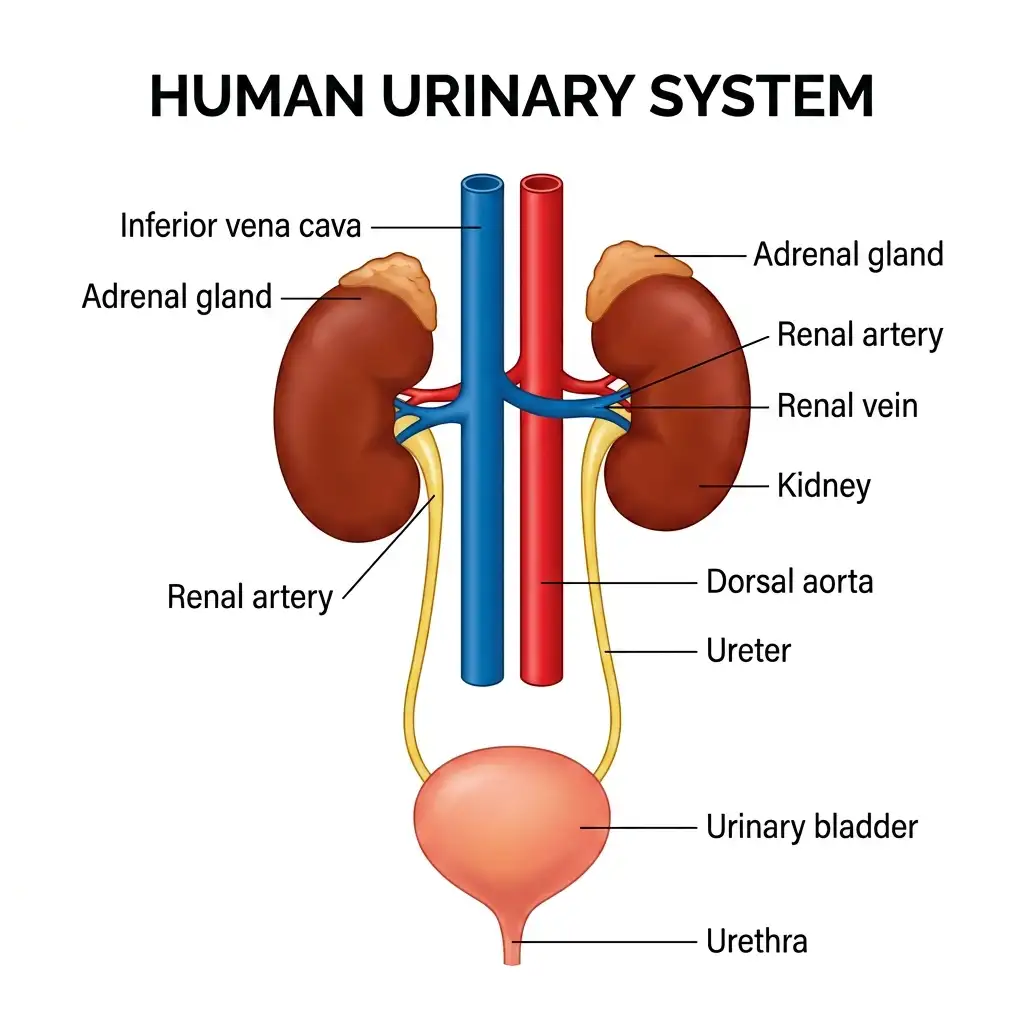
The Human Urinary System: Comprises a pair of kidneys, two ureters, a urinary bladder, and a urethra, responsible for filtering wastes from the blood.
🔬 Detailed Anatomical Description
The human excretory system consists of: (1) A pair of reddish-brown, bean-shaped kidneys located in the abdominal cavity; (2) Adrenal glands cap each kidney; (3) Major blood vessels: the dorsal aorta supplying oxygenated blood via renal arteries, and the inferior vena cava draining filtered blood via renal veins; (4) A pair of thin, muscular ureters; (5) A stretchable urinary bladder for temporary urine storage; and (6) The urethra, through which urine is expelled during micturition.
B KIDNEYS — POSITION AND EXTERNAL FEATURES
| FEATURE | DETAIL |
|---|---|
| Shape | Bean-shaped ⭐⭐ |
| Colour | Reddish brown ⭐ |
| Location | Between last thoracic and third lumbar vertebra, close to dorsal inner wall of abdominal cavity ⭐⭐⭐ |
| Size | 10–12 cm length, 5–7 cm width, 2–3 cm thickness ⭐⭐ |
| Weight | About 120–170 g ⭐ |
| Hilum | Inner concave notch through which ureter, blood vessels and nerves enter ⭐⭐⭐ |
| Renal pelvis | Funnel-shaped cavity inside hilum ⭐⭐ |
| Calyces | Projections of renal pelvis ⭐⭐ |
| Outer covering | Tough capsule ⭐ |
C KIDNEY INTERNAL STRUCTURE
| PART | DETAIL |
|---|---|
| Outer zone | Cortex ⭐⭐⭐ |
| Inner zone | Medulla ⭐⭐⭐ |
| Medulla divided into | Medullary pyramids projecting into calyces ⭐⭐⭐ |
| Cortex between pyramids | Renal columns / Columns of Bertini ⭐⭐⭐ |
NEET 2023: Outer part = cortex, inner part = medulla.
Kidney Longitudinal Section
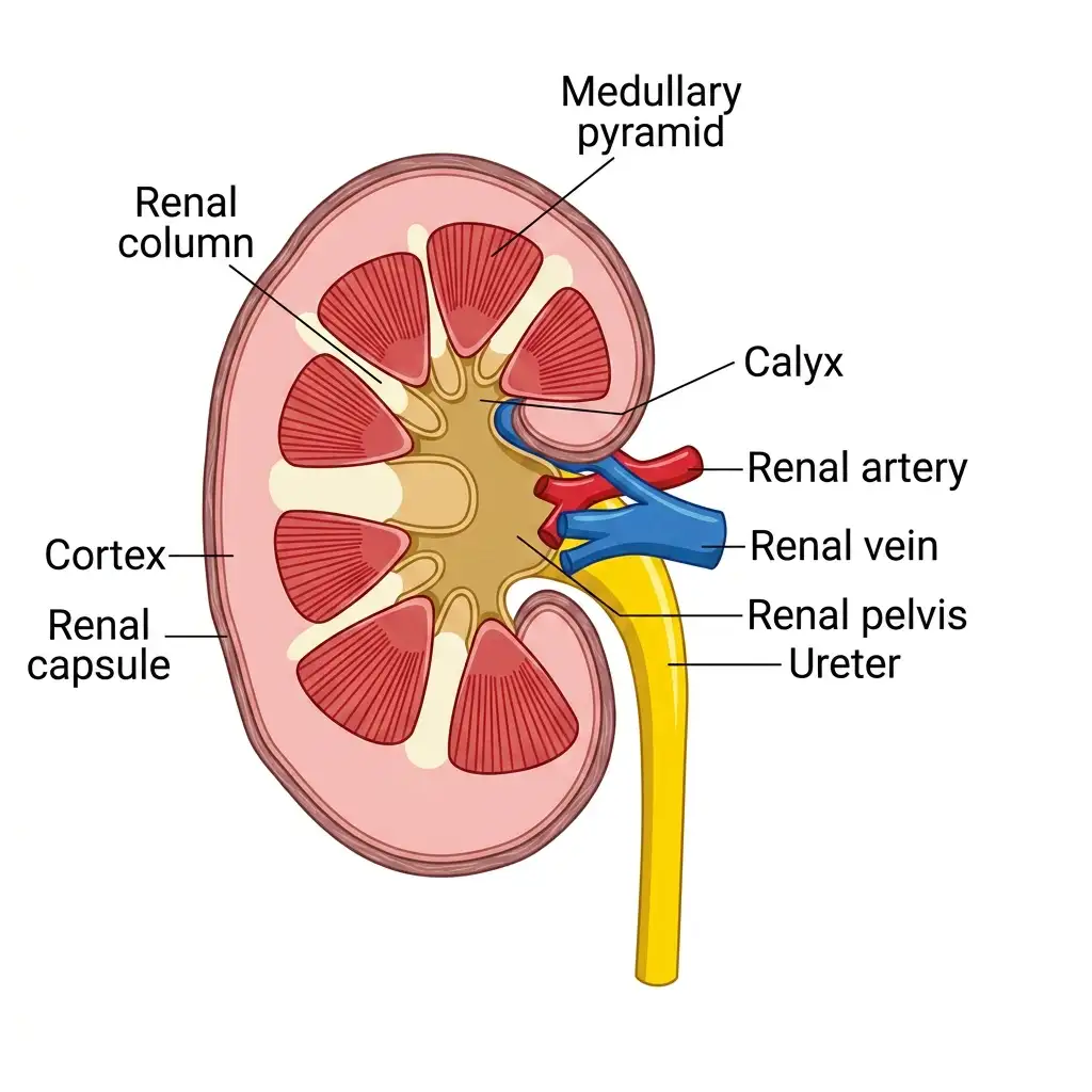
Longitudinal Section of a Kidney: Reveals the outer cortex, inner medulla containing medullary pyramids, calyces, and the funnel-shaped renal pelvis.
🔬 Detailed Anatomical Description
A diagrammatic longitudinal section reveals the highly compartmentalized internal kidney anatomy. The outer layer is a tough, fibrous renal capsule. Internally, the kidney is divided into an outer cortex and an inner medulla. The medulla is split into several conical medullary pyramids projecting into cup-like calyces. Calyces drain into a wide central funnel-shaped space called the renal pelvis, which leads directly into the ureter. The cortex extends between pyramids as renal columns of Bertin.
D EXTRA HIGH-YIELD ASSOCIATION
| FEATURE | DETAIL |
|---|---|
| Adrenal gland location | At the anterior part / top of kidney ⭐ |
| Hormone type | Catecholamines — stimulate glycogen breakdown ⭐ |
NEET 2013: Adrenal gland association with kidney.
V. NEPHRON — FUNCTIONAL UNIT OF KIDNEY
| FEATURE | DETAIL |
|---|---|
| Functional unit of kidney | Nephron ⭐⭐⭐ |
| Number per kidney | Nearly 1 million ⭐⭐⭐ |
A TWO MAIN PARTS OF A NEPHRON
| PART | DETAIL |
|---|---|
| Glomerulus | Tuft of capillaries formed by afferent arteriole ⭐⭐⭐ |
| Blood leaves glomerulus by | Efferent arteriole ⭐⭐⭐ |
| Renal tubule | Begins with Bowman’s capsule ⭐⭐⭐ |
B MALPIGHIAN BODY / RENAL CORPUSCLE
| FEATURE | DETAIL |
|---|---|
| Bowman’s capsule | Double-walled cup-like structure enclosing glomerulus ⭐⭐⭐ |
| Glomerulus + Bowman’s capsule | Malpighian body / Renal corpuscle ⭐⭐⭐ |
C SEQUENCE OF TUBULE
| SEGMENT | FULL FORM |
|---|---|
| PCT | Proximal convoluted tubule |
| DCT | Distal convoluted tubule |
| CT | Collecting tubule / collecting duct |
Nephron Tubules and Vessels
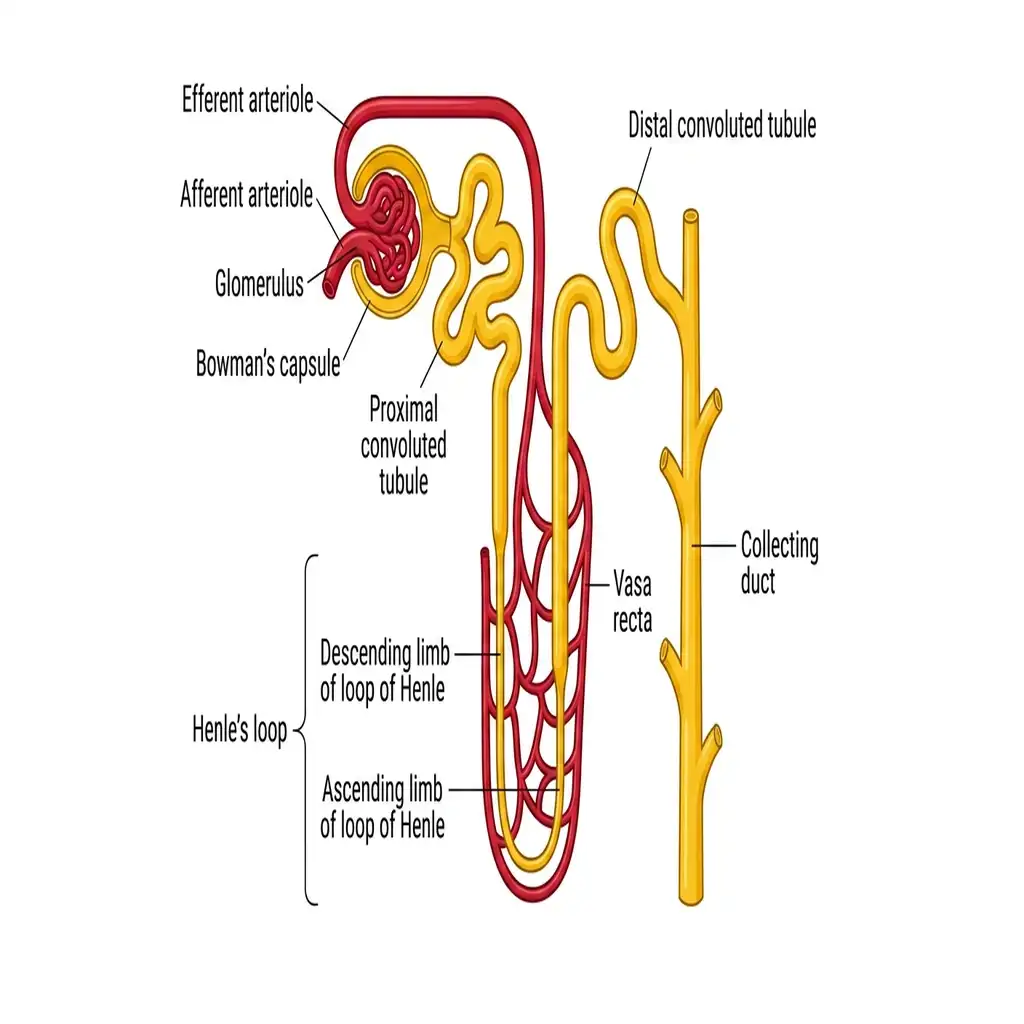
The Nephron and Vasa Recta: The structural unit of filtration, displaying the proximal/distal tubules closely intertwined with the blood vessels.
🔬 Detailed Anatomical Description
A nephron is the functional unit of the kidney, consisting of a renal corpuscle and a renal tubule. The tubule begins at Bowman's capsule, twists into the proximal convoluted tubule (PCT), dips into the medulla as the loop of Henle (with descending and ascending limbs), and twists again into the distal convoluted tubule (DCT), which empties into the collecting duct. The vascular network comprises the afferent arteriole feeding the glomerulus, the efferent arteriole exiting the capsule, and the vasa recta loop surrounding the loop of Henle.
D REGION-WISE LOCATION
| STRUCTURE | REGION |
|---|---|
| Malpighian corpuscle | Cortex ⭐⭐⭐ |
| PCT | Cortex ⭐⭐⭐ |
| DCT | Cortex ⭐⭐⭐ |
| Loop of Henle | Dips into medulla ⭐⭐⭐ |
VI. TYPES OF NEPHRONS
| TYPE | DETAIL |
|---|---|
| Cortical nephrons | Majority; loop of Henle is short and extends very little into medulla ⭐⭐⭐ |
| Juxtamedullary nephrons | Fewer; loop of Henle is long and extends deep into medulla ⭐⭐⭐ |
| ADDITIONAL FEATURE | DETAIL |
|---|---|
| Vasa recta in cortical nephron | Absent or highly reduced ⭐⭐⭐ |
NEET 2023, 2024, 2026: Juxtamedullary nephron = long loop of Henle.
NEET 2023, 2026: Cortical nephron = short loop of Henle.
NEET 2023, 2024, 2026 context: Cortical nephrons have short loops of Henle; juxtamedullary nephrons have long loops of Henle that extend deep into the medulla.
VII. BLOOD VESSELS OF NEPHRON
| FEATURE | DETAIL |
|---|---|
| Efferent arteriole forms | Fine capillary network around tubule = Peritubular capillaries ⭐⭐⭐ |
| Minute vessels parallel to Henle’s loop | Vasa recta ⭐⭐⭐ |
| Shape of vasa recta | U-shaped ⭐⭐ |
VIII. URINE FORMATION — OVERVIEW
IX. GLOMERULAR FILTRATION
| FEATURE | DETAIL |
|---|---|
| Definition | Filtration of blood by glomerulus ⭐⭐⭐ |
| Blood filtered by kidneys/min | 1100–1200 mL/min ⭐⭐⭐ |
| This is about | 1/5th of blood pumped by each ventricle per minute ⭐⭐ |
| Driving force | Glomerular capillary blood pressure ⭐⭐⭐ |
Filtration Barrier — 3 Layers ⭐⭐⭐
| LAYER | DETAIL |
|---|---|
| 1 | Endothelium of glomerular blood vessels ⭐⭐⭐ |
| 2 | Basement membrane ⭐⭐⭐ |
| 3 | Epithelium of Bowman’s capsule ⭐⭐⭐ |
Podocytes ⭐⭐⭐
| FEATURE | DETAIL |
|---|---|
| Bowman’s capsule epithelium cells | Podocytes ⭐⭐⭐ |
| Podocytes leave | Minute spaces called filtration slits / slit pores ⭐⭐⭐ |
Malpighian Body Structure
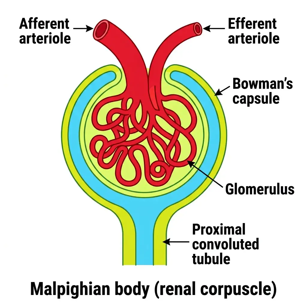
Structure of a Malpighian Body: Composed of the glomerulus capillary tuft and its enveloping Bowman's capsule, initiating ultrafiltration.
🔬 Detailed Anatomical Description
The Malpighian body (renal corpuscle) is the site where blood filtration begins. It is formed by: (1) The glomerulus, a tuft of capillaries receiving blood from a wide afferent arteriole and draining into a narrower efferent arteriole, creating high hydrostatic pressure; and (2) Bowman's capsule, a double-walled epithelial cup that wraps around the glomerulus. The inner visceral wall contains podocytes with filtration slits that allow water and small solutes to pass into the capsular lumen (ultrafiltration).
Nature of Filtrate ⭐⭐⭐
| FEATURE | DETAIL |
|---|---|
| Filtration type | Ultrafiltration ⭐⭐⭐ |
| What passes into Bowman’s capsule? | Almost all plasma constituents except proteins ⭐⭐⭐ |
| Fluid filtered | Protein-free filtrate ⭐⭐⭐ |
GFR ⭐⭐⭐
| FEATURE | DETAIL |
|---|---|
| GFR = | Glomerular Filtration Rate ⭐⭐⭐ |
| Definition | Volume of filtrate formed per minute ⭐⭐⭐ |
| Normal value | 125 mL/min ⭐⭐⭐ |
| Per day | 180 L/day ⭐⭐⭐ |
NEET 2011: Endothelium is part of filtration membrane.
NEET 2023: Malpighian corpuscle = site of ultrafiltration.
X. JUXTA GLOMERULAR APPARATUS (JGA)
| FEATURE | DETAIL |
|---|---|
| Formed by | Cellular modifications of DCT and afferent arteriole at point of contact ⭐⭐⭐ |
| Function | Regulation of GFR ⭐⭐⭐ |
| If GFR falls | JG cells release renin ⭐⭐⭐ |
| Effect | Renin restores glomerular blood flow and GFR toward normal ⭐⭐ |
NEET 2020, 2023, 2026 (reinforced): Fall in GFR activates the juxtaglomerular apparatus (JGA) which releases renin.
XI. REABSORPTION
| FEATURE | DETAIL |
|---|---|
| Filtrate formed/day | 180 L ⭐⭐⭐ |
| Urine excreted/day | ~1.5 L ⭐⭐⭐ |
| Therefore | Nearly 99% of filtrate is reabsorbed ⭐⭐⭐ |
Active vs Passive Reabsorption ⭐⭐⭐
| TYPE | SUBSTANCES |
|---|---|
| Active | Glucose, Amino acids, Na⁺ ⭐⭐⭐ |
| Passive | Water, nitrogenous wastes ⭐⭐⭐ |
XII. SELECTIVE SECRETION
| FEATURE | DETAIL |
|---|---|
| Secreted into filtrate | H⁺, K⁺, NH₃ (ammonia) ⭐⭐⭐ |
| Importance | Maintains ionic balance and acid-base balance ⭐⭐⭐ |
XIII. FUNCTION OF DIFFERENT PARTS OF TUBULE
Nephron Reabsorption & Secretion
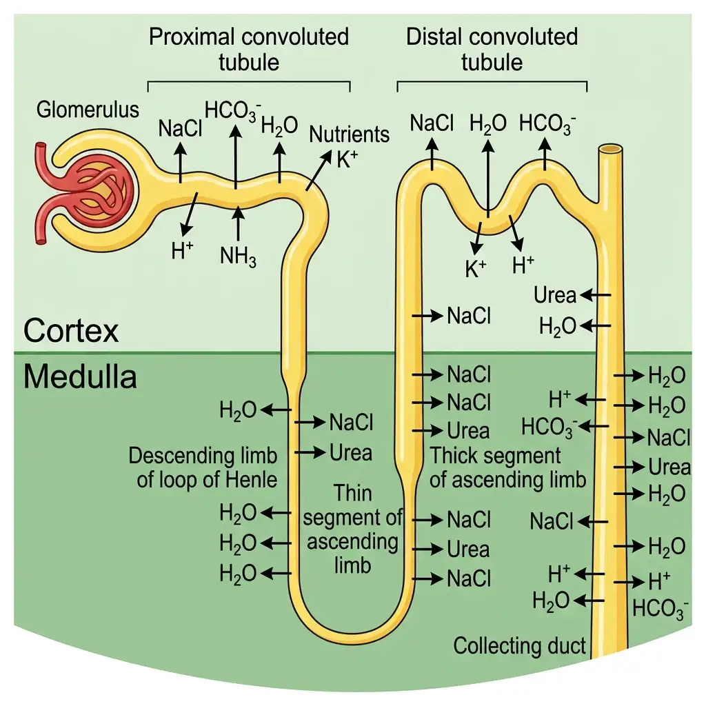
Tubular Reabsorption and Secretion: Illustrates active and passive transport of water, salts, nutrients, and waste ions in different nephron zones.
🔬 Detailed Anatomical Description
Filtrate is processed through selective reabsorption and tubular secretion. (1) PCT: Reabsorbs 70-80% of electrolytes and water, glucose, and amino acids; secretes H+, NH3, and K+ to maintain pH. (2) Descending limb of Henle: Permeable to water but impermeable to salts, concentrating the filtrate. (3) Ascending limb: Impermeable to water but active/passive transport of NaCl dilutes the filtrate. (4) DCT: Conditional reabsorption of Na+ and water; secretes K+ and H+. (5) Collecting duct: Reabsorbs water and some urea.
A PROXIMAL CONVOLUTED TUBULE (PCT)
| FEATURE | DETAIL |
|---|---|
| Lining | Simple cuboidal brush border epithelium ⭐⭐⭐ |
| Brush border role | Increases surface area for reabsorption ⭐⭐⭐ |
| Reabsorbs | Nearly all essential nutrients and 70–80% electrolytes and water ⭐⭐⭐ |
| Selective secretion | H⁺ and NH₃ into filtrate ⭐⭐⭐ |
| Also reabsorbs | HCO₃⁻ ⭐⭐⭐ |
| Role | Maintains pH and ionic balance ⭐⭐ |
NTA trap: PCT is NOT simple columnar — it is simple cuboidal brush border epithelium.
NEET 2016: PCT actively reabsorbs Na⁺.
NEET 2015: Removal of PCT → more dilute urine.
B HENLE’S LOOP
Descending Limb ⭐⭐⭐
| FEATURE | DETAIL |
|---|---|
| Permeable to | Water ⭐⭐⭐ |
| Almost impermeable to | Electrolytes ⭐⭐⭐ |
| Effect | Filtrate becomes more concentrated as it moves down ⭐⭐ |
Ascending Limb ⭐⭐⭐
| FEATURE | DETAIL |
|---|---|
| Reabsorption of water | Absent / minimum ⭐⭐⭐ |
| Impermeable to | Water ⭐⭐⭐ |
| Allows transport of | Electrolytes actively or passively ⭐⭐⭐ |
| Effect | Efflux of electrolytes causes dilution of filtrate ⭐⭐⭐ |
Overall role ⭐⭐⭐
| FEATURE | DETAIL |
|---|---|
| Main function | Helps maintain high osmolarity of medullary interstitial fluid ⭐⭐⭐ |
NEET 2017, 2024: Descending limb = water permeable.
NEET 2017, 2023: Ascending limb = water impermeable, electrolyte transport.
C DISTAL CONVOLUTED TUBULE (DCT)
| FEATURE | DETAIL |
|---|---|
| Conditional reabsorption | Na⁺ and water ⭐⭐⭐ |
| Also reabsorbs | HCO₃⁻ ⭐⭐⭐ |
| Selective secretion | H⁺, K⁺, NH₃ ⭐⭐⭐ |
| Role | Maintains pH and Na⁺–K⁺ balance in blood ⭐⭐⭐ |
NEET 2016: Conditional Na⁺ and water reabsorption occurs in DCT.
D COLLECTING DUCT
| FEATURE | DETAIL |
|---|---|
| Water reabsorption | Large amount of water reabsorbed ⭐⭐⭐ |
| Function | Produces concentrated urine ⭐⭐⭐ |
| Urea movement | Allows small amount of urea into medullary interstitium ⭐⭐⭐ |
| Why? | Helps maintain osmolarity of medulla ⭐⭐⭐ |
| Also helps in | pH and ionic balance by secretion of H⁺ and K⁺ ⭐⭐ |
XIV. MECHANISM OF CONCENTRATION OF URINE
A COUNTER CURRENT MECHANISM
| FEATURE | DETAIL |
|---|---|
| Main structures involved | Henle’s loop and vasa recta ⭐⭐⭐ |
| Why important? | Mammals produce concentrated urine using this mechanism ⭐⭐⭐ |
| Basis | Close proximity + opposite direction of flow ⭐⭐⭐ |
Counter-Current Mechanism
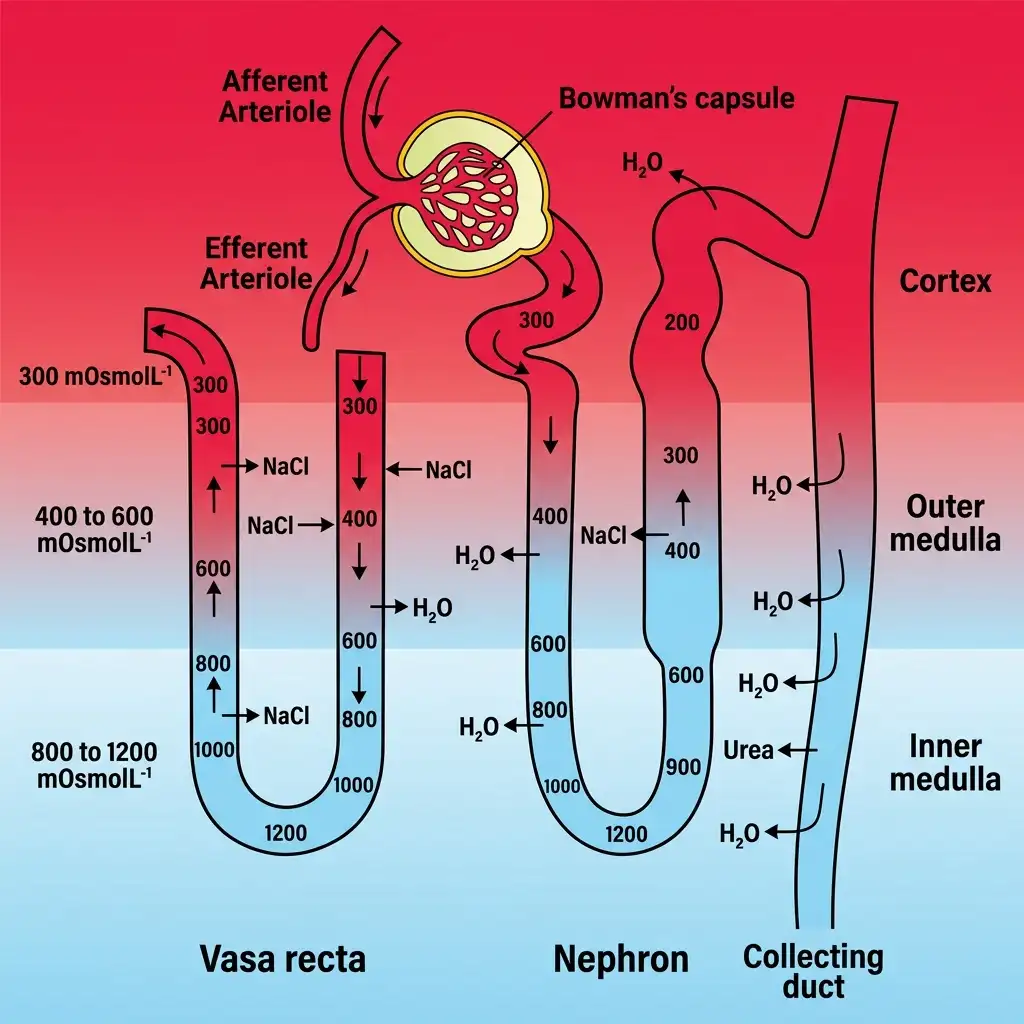
The Counter-Current Mechanism: Maintained by the loop of Henle and vasa recta to establish a high osmolarity gradient in the medullary interstitium.
🔬 Detailed Anatomical Description
The kidney concentrates urine using a counter-current mechanism involving the loop of Henle and vasa recta. (1) Opposing flow directions in limbs create a multiplier effect. (2) Active transport of NaCl out of the ascending limb and passive diffusion of urea from the collecting duct create a high medullary interstitial osmolarity (from 300 mOsmol/L in cortex up to 1200 mOsmol/L in inner medulla). (3) This gradient draws water out of the descending limb and collecting duct, concentrating urine up to 4x plasma osmolarity.
B COUNTER CURRENT PATTERN
| STRUCTURE | DIRECTION |
|---|---|
| Henle’s loop | Filtrate flows in opposite directions in two limbs ⭐⭐⭐ |
| Vasa recta | Blood flows in opposite directions in two limbs ⭐⭐⭐ |
C OSMOTIC GRADIENT
| REGION | OSMOLARITY |
|---|---|
| Cortex | 300 mOsmol L⁻¹ ⭐⭐⭐ |
| Inner medulla | 1200 mOsmol L⁻¹ ⭐⭐⭐ |
| FEATURE | DETAIL |
|---|---|
| Gradient caused mainly by | NaCl and Urea ⭐⭐⭐ |
| NaCl transported by | Ascending limb of Henle’s loop ⭐⭐⭐ |
| Urea recycled via | Thin ascending limb + collecting duct ⭐⭐ |
D RESULT
| FEATURE | DETAIL |
|---|---|
| Counter current mechanism maintains | Medullary interstitial concentration gradient ⭐⭐⭐ |
| This helps in | Easy movement of water from collecting duct ⭐⭐⭐ |
| Final result | Formation of concentrated urine ⭐⭐⭐ |
| Human kidney can concentrate urine | Nearly 4 times more than initial filtrate ⭐⭐⭐ |
NEET 2020: Counter current works due to close proximity of Henle’s loop and vasa recta.
NEET 2018, 2019, 2024: Hyperosmolarity of inner medulla is essential for concentrated urine.
XV. REGULATION OF KIDNEY FUNCTION
XVI. ADH / VASOPRESSIN
| FEATURE | DETAIL |
|---|---|
| Full form | Antidiuretic hormone ⭐⭐⭐ |
| Also called | Vasopressin ⭐⭐ |
| Released from | Neurohypophysis / Posterior pituitary ⭐⭐⭐ |
| Trigger | Excessive fluid loss activates osmoreceptors → hypothalamus stimulates ADH release ⭐⭐⭐ |
| Action | Facilitates water reabsorption from later parts of tubule ⭐⭐⭐ |
| Result | Prevents diuresis ⭐⭐⭐ |
| Additional effect | Acts as vasoconstrictor → increases blood pressure ⭐⭐⭐ |
NEET 2022, 2024: ADH released from neurohypophysis, not adenohypophysis.
NEET 2023, 2024: ADH prevents diuresis.
NEET 2023: ADH increases BP by vasoconstriction.
NEET 2022, 2023, 2024, 2026 context: ADH (vasopressin) is released from the posterior pituitary (neurohypophysis) and promotes water reabsorption to prevent diuresis.
XVII. RENIN–ANGIOTENSIN–ALDOSTERONE SYSTEM (RAAS)
A TRIGGER
| FEATURE | DETAIL |
|---|---|
| Trigger | Fall in glomerular blood flow, glomerular blood pressure, or GFR ⭐⭐⭐ |
B SEQUENCE
C ACTIONS
| COMPONENT | ACTION |
|---|---|
| Renin | Starts RAAS ⭐⭐⭐ |
| Angiotensin II | Powerful vasoconstrictor; increases glomerular blood pressure and GFR ⭐⭐⭐ |
| Aldosterone | Causes reabsorption of Na⁺ and water from distal tubule / DCT ⭐⭐⭐ |
| Final outcome | ↑ Blood pressure and ↑ GFR ⭐⭐⭐ |
NEET 2014, 2022: Aldosterone increases Na⁺ reabsorption.
NEET 2020, 2023: Fall in GFR triggers renin release.
NEET 2023: Full RAAS sequence is high-yield.
NEET 2014, 2020, 2022, 2023, 2026 context: Fall in blood pressure or GFR triggers renin release from JGA, leading to angiotensin II formation and aldosterone release.
XVIII. ATRIAL NATRIURETIC FACTOR (ANF)
| FEATURE | DETAIL |
|---|---|
| Released by | Atria of heart ⭐⭐⭐ |
| Trigger | Increased blood flow / stretch of atria ⭐⭐⭐ |
| Action | Vasodilation ⭐⭐⭐ |
| Effect | Decreases blood pressure ⭐⭐⭐ |
| Role | Acts as a check on RAAS ⭐⭐⭐ |
NEET 2023: ANF released when atrial blood flow increases.
NEET 2017: ANF opposes renin-angiotensin mechanism.
TRAP: Low blood pressure does not stimulate ANF release.
XIX. DIURETICS
| SUBSTANCE | EFFECT |
|---|---|
| Alcohol | Promotes large quantity of dilute urine ⭐⭐ |
| Caffeine | Same ⭐⭐ |
| ANF | Same ⭐⭐ |
| Renin | Does not act as diuretic; helps retain water/Na⁺ ⭐⭐ |
NEET 2015: Alcohol, caffeine, ANF are diuretic-type factors.
XX. MICTURITION
| FEATURE | DETAIL |
|---|---|
| Urine stored in | Urinary bladder ⭐⭐⭐ |
| Stored until | Voluntary signal from CNS ⭐⭐⭐ |
| Trigger | Bladder wall stretches as urine accumulates ⭐⭐⭐ |
| Stretch receptors | Send signals to CNS ⭐⭐ |
| CNS response | Causes contraction of bladder smooth muscle + relaxation of urethral sphincter ⭐⭐⭐ |
| Micturition | Release of urine ⭐⭐⭐ |
| Micturition reflex | Neural mechanism causing urination ⭐⭐⭐ |
Urine Facts ⭐⭐⭐
| FEATURE | DETAIL |
|---|---|
| Urine output/day | 1 to 1.5 L/day ⭐⭐⭐ |
| Nature | Light yellow, watery fluid, characteristic odour ⭐ |
| pH | Slightly acidic, about 6.0 ⭐⭐⭐ |
| Urea excreted/day | 25–30 g/day ⭐⭐⭐ |
NEET 2015, 2026 context: Human urine is acidic (pH ~6.0) because H⁺ is actively secreted into filtrate. Micturition is the process of urination controlled by a neural reflex involving stretch receptors in the bladder wall.
XXI. URINE ABNORMALITIES
| CONDITION | MEANING | INDICATES |
|---|---|---|
| Glycosuria | Glucose in urine ⭐⭐⭐ | Diabetes mellitus ⭐⭐⭐ |
| Ketonuria | Ketone bodies in urine ⭐⭐⭐ | Diabetes mellitus ⭐⭐⭐ |
NEET 2018: Glycosuria + ketonuria = diabetes mellitus.
XXII. ROLE OF OTHER ORGANS IN EXCRETION
A LUNGS
| FEATURE | DETAIL |
|---|---|
| Remove | Large amount of CO₂ and water ⭐⭐⭐ |
| CO₂ removed | About 200 mL/min ⭐⭐⭐ |
B LIVER
| FEATURE | DETAIL |
|---|---|
| Bile contains | Bilirubin, biliverdin, cholesterol, degraded steroid hormones, vitamins, drugs ⭐⭐⭐ |
| Special PYQ fact | Hepatic vein carries largest amount of urea in mammals ⭐⭐ |
NEET 2016: Hepatic vein = largest urea carrier.
C SKIN
| STRUCTURE | SECRETION | EXCRETORY SUBSTANCES |
|---|---|---|
| Sweat glands | Sweat | NaCl, small amount of urea, lactic acid ⭐⭐⭐ |
| Sebaceous glands | Sebum | Sterols, hydrocarbons, waxes ⭐⭐⭐ |
| ADDITIONAL NOTE | DETAIL |
|---|---|
| Sweat primary role | Cooling effect, but also excretory ⭐ |
| Sebum | Gives protective oily covering to skin ⭐ |
XXIII. DISORDERS OF EXCRETORY SYSTEM
A UREMIA
| FEATURE | DETAIL |
|---|---|
| Definition | Accumulation of urea in blood ⭐⭐⭐ |
| Effect | Very harmful; may lead to kidney failure ⭐⭐⭐ |
| Treatment | Hemodialysis ⭐⭐⭐ |
B HEMODIALYSIS
| FEATURE | DETAIL |
|---|---|
| Artificial kidney | Dialysing unit used in hemodialysis ⭐⭐⭐ |
| Blood source | Drawn from a convenient artery ⭐⭐ |
| Anticoagulant added | Heparin ⭐⭐⭐ |
| Dialysis tube | Coiled cellophane tube ⭐⭐⭐ |
| Dialysing fluid composition | Same as plasma except nitrogenous wastes ⭐⭐⭐ |
| Principle | Diffusion along concentration gradient across porous membrane ⭐⭐ |
| Blood returned to body | Through vein after adding anti-heparin ⭐⭐⭐ |
C HEMODIALYSIS LIMITATION
| FEATURE | DETAIL |
|---|---|
| Artificial kidney does NOT replace | Endocrine functions of kidney ⭐⭐ |
| Therefore patient may develop | ↓ RBC production (lack of erythropoietin) + ↓ calcium absorption from GIT (lack of active vitamin D) ⭐⭐ |
NEET 2019, 2026 context: Hemodialysis is used in cases of uremia or kidney failure. It does not replace the endocrine functions of the kidney.
D KIDNEY TRANSPLANTATION
| FEATURE | DETAIL |
|---|---|
| Ultimate correction for | Acute renal failure / kidney failure ⭐⭐⭐ |
| Preferred donor | Close relative to reduce rejection ⭐⭐ |
E RENAL CALCULI
| FEATURE | DETAIL |
|---|---|
| Definition | Stone / insoluble mass of crystallised salts (especially oxalates) in kidney ⭐⭐⭐ |
F GLOMERULONEPHRITIS
| FEATURE | DETAIL |
|---|---|
| Definition | Inflammation of glomeruli of kidney ⭐⭐⭐ |
G GOUT
| FEATURE | DETAIL |
|---|---|
| Cause | Accumulation of uric acid crystals in joints ⭐⭐ |
| Result | Inflammation of joints ⭐⭐ |
NEET 2018: Disorder matching — glycosuria, gout, renal calculi, glomerulonephritis.
XXIV. RAPID REVISION — KEY COMPARISON TABLES
TABLE 1: Nitrogenous Wastes ⭐⭐⭐
| WASTE | TOXICITY | WATER REQUIRED |
|---|---|---|
| Ammonia | Most toxic | Maximum |
| Urea | Moderately toxic | Moderate |
| Uric acid | Least toxic | Minimum |
TABLE 2: Animal Types ⭐⭐⭐
| TYPE | PRODUCT | EXAMPLES |
|---|---|---|
| Ammonotelic | Ammonia | Bony fishes, aquatic amphibians, aquatic insects |
| Ureotelic | Urea | Mammals, terrestrial amphibians, marine fishes |
| Uricotelic | Uric acid | Reptiles, birds, land snails, insects |
TABLE 3: Excretory Structures in Animals ⭐⭐⭐
| STRUCTURE | ORGANISM |
|---|---|
| Flame cells / Protonephridia | Platyhelminthes, rotifers, some annelids, Amphioxus |
| Nephridia | Earthworm, annelids |
| Malpighian tubules | Insects |
| Green glands | Prawns / crustaceans |
| Kidneys | Vertebrates |
TABLE 4: Kidney Internal Parts ⭐⭐⭐
| PART | DETAIL |
|---|---|
| Cortex | Outer zone |
| Medulla | Inner zone |
| Medullary pyramids | Conical masses in medulla |
| Columns of Bertini | Cortex between pyramids |
| Calyces | Receive projections of pyramids |
| Renal pelvis | Funnel-shaped cavity |
TABLE 5: Nephron Structure ⭐⭐⭐
| PART | DETAIL |
|---|---|
| Glomerulus | Tuft of capillaries |
| Bowman’s capsule | Double-walled cup |
| Malpighian body | Glomerulus + Bowman’s capsule |
| PCT | Major reabsorption |
| Loop of Henle | Osmotic gradient |
| DCT | Conditional reabsorption |
| Collecting duct | Concentrated urine formation |
TABLE 6: Cortical vs Juxtamedullary Nephrons ⭐⭐⭐
| FEATURE | CORTICAL | JUXTAMEDULLARY |
|---|---|---|
| Number | Majority | Few |
| Loop of Henle | Short | Long |
| Extends into medulla | Very little | Deep |
| Vasa recta | Absent / reduced | Present |
TABLE 7: Urine Formation ⭐⭐⭐
| STEP | DETAIL |
|---|---|
| Glomerular filtration | Blood filtered in glomerulus |
| Reabsorption | Useful substances taken back |
| Selective secretion | H⁺, K⁺, NH₃ secreted |
TABLE 8: Tubular Functions ⭐⭐⭐
| SEGMENT | MAIN FUNCTION |
|---|---|
| PCT | Reabsorbs nutrients, 70–80% water / electrolytes; secretes H⁺, NH₃ |
| Descending Henle | Water reabsorption |
| Ascending Henle | Electrolyte transport; impermeable to water |
| DCT | Conditional Na⁺, water reabsorption; secretes H⁺, K⁺, NH₃ |
| Collecting duct | Concentrated urine; urea recycling |
TABLE 9: Hormonal Control ⭐⭐⭐
| HORMONE / FACTOR | SOURCE | MAIN EFFECT |
|---|---|---|
| ADH / Vasopressin | Neurohypophysis | Water reabsorption |
| Renin | JG cells | Starts RAAS |
| Angiotensin II | Blood pathway product | Vasoconstriction |
| Aldosterone | Adrenal cortex | Na⁺ + water reabsorption |
| ANF | Atria of heart | Vasodilation, ↓ BP |
TABLE 10: Disorders ⭐⭐⭐
| DISORDER | MEANING |
|---|---|
| Uremia | Urea accumulation in blood |
| Glycosuria | Glucose in urine |
| Ketonuria | Ketone bodies in urine |
| Renal calculi | Kidney stones |
| Glomerulo-nephritis | Inflammation of glomeruli |
| Gout | Uric acid crystal deposition in joints |
XXV. KEY NUMBERS — QUICK REFERENCE
| PARAMETER | VALUE |
|---|---|
| Blood filtered by kidneys/min | 1100–1200 mL |
| GFR | 125 mL/min |
| Filtrate/day | 180 L/day |
| Urine/day | 1–1.5 L/day |
| Reabsorption | ~99% |
| Kidney size | 10–12 × 5–7 × 2–3 cm |
| Kidney weight | 120–170 g |
| Nephrons/kidney | ~1 million |
| Osmolarity in cortex | 300 mOsmol L⁻¹ |
| Osmolarity in inner medulla | 1200 mOsmol L⁻¹ |
| Urine concentration ability | 4× initial filtrate |
| Urine pH | 6.0 |
| Urea/day | 25–30 g/day |
| CO₂ removed by lungs | 200 mL/min |
XXVI. COMMON EXAM TRAPS — QUICK REFERENCE
| TRAP / QUESTION | CORRECT ANSWER |
|---|---|
| Most toxic nitrogenous waste? | Ammonia |
| Least toxic nitrogenous waste? | Uric acid |
| Birds are ureotelic? | No, uricotelic |
| Pavo excretes nitrogenous waste in? | Pellet / paste |
| Flame cells found in Amphioxus? | Yes |
| Functional unit of kidney? | Nephron |
| Malpighian body = ? | Glomerulus + Bowman’s capsule |
| Ultrafiltration site? | Malpighian corpuscle |
| PCT epithelium columnar? | False — simple cuboidal brush border |
| Descending limb permeable to? | Water |
| Ascending limb permeable to water? | No |
| Ascending limb allows transport of? | Electrolytes |
| DCT reabsorbs? | Na⁺, water conditionally, HCO₃⁻ |
| Urine acidic because? | H⁺ secretion into filtrate |
| JGA formed by? | DCT + afferent arteriole |
| Fall in GFR causes release of? | Renin |
| ADH released from adenohypophysis? | No, from neurohypophysis |
| ADH promotes diuresis? | No, prevents diuresis |
| ANF released in low blood pressure? | No |
| ANF effect? | Vasodilation, ↓ BP |
| Aldosterone effect? | ↑ Na⁺ and water reabsorption |
| Dialysing fluid contains nitrogenous wastes? | No |
| Artificial kidney replaces endocrine functions? | No |
| Renal calculi means? | Crystallised salts / kidney stones |
| Glomerulo-nephritis means? | Inflammation of glomeruli |
| Glycosuria + ketonuria indicate? | Diabetes mellitus |
| Hepatic vein carries what? | Largest amount of urea |