I. INTRODUCTION & BASICS
| FEATURE | DETAIL |
|---|---|
| Movement | Significant feature of living beings ⭐⭐ |
| Locomotion | Voluntary movement causing change of place / location ⭐⭐⭐ |
| Key relation | All locomotions are movements, but all movements are not locomotions ⭐⭐⭐ |
| Therefore | Movement and locomotion cannot be studied separately ⭐⭐⭐ |
Examples showing movement ≠ locomotion ⭐⭐
| ORGANISM / STRUCTURE | ROLE |
|---|---|
| Paramoecium cilia | Move food through cytopharynx + locomotion ⭐ |
| Hydra tentacles | Capture prey + locomotion ⭐ |
| Human limbs | Change body posture + locomotion ⭐ |
Why animals move / locomote ⭐⭐
| PURPOSE | DETAIL |
|---|---|
| Search of food | ⭐ |
| Shelter | ⭐ |
| Mate | ⭐ |
| Suitable breeding grounds | ⭐ |
| Favourable climatic conditions | ⭐ |
| Escape from enemies / predators | ⭐ |
II. TYPES OF MOVEMENT
A MAIN TYPES OF MOVEMENT IN HUMAN BODY
| TYPE | EXAMPLE | MECHANISM / SITE |
|---|---|---|
| Amoeboid movement | Macrophages, Leucocytes (WBCs) ⭐⭐⭐ | By pseudopodia; involves microfilaments ⭐⭐ |
| Ciliary movement | Trachea, passage of ova through female reproductive tract ⭐⭐⭐ | Coordinated beating of cilia ⭐⭐ |
| Muscular movement | Movement of limbs, jaws, tongue, eyelids etc. ⭐⭐⭐ | Due to contraction of muscles ⭐⭐⭐ |
B FLAGELLAR MOVEMENT
| EXAMPLE | ROLE |
|---|---|
| Spermatozoa | Swimming ⭐⭐⭐ |
| Sponges | Maintenance of water current in canal system ⭐⭐ |
| Protists / Protozoans (e.g. Euglena) | Locomotion ⭐⭐ |
In human body, the three main movement types are amoeboid, ciliary and muscular; flagellar movement is a special example seen in spermatozoa.
III. MUSCLE — BASIC FACTS
| FEATURE | DETAIL |
|---|---|
| Muscle tissue origin | Mesodermal ⭐⭐⭐ |
| Contribution to body weight | About 40–50% of body weight in adult human ⭐⭐⭐ |
| Properties of muscles | Excitability, Contractility, Extensibility, Elasticity ⭐⭐⭐ |
IV. TYPES OF MUSCLES
| FEATURE | SKELETAL MUSCLE | VISCERAL (SMOOTH) MUSCLE | CARDIAC MUSCLE |
|---|---|---|---|
| Other name | Striated muscle ⭐⭐⭐ | Smooth / Unstriated / Nonstriated ⭐⭐⭐ | Striated muscle ⭐⭐ |
| Control | Voluntary ⭐⭐⭐ | Involuntary ⭐⭐⭐ | Involuntary ⭐⭐⭐ |
| Location | Attached to skeletal elements ⭐⭐⭐ | Wall of internal hollow organs ⭐⭐⭐ | Only in heart ⭐⭐⭐ |
| Main role | Locomotion and posture ⭐⭐⭐ | Movement of food / gametes etc. ⭐⭐ | Rhythmic contraction of heart ⭐⭐ |
| Appearance | Striped ⭐⭐ | Smooth ⭐⭐ | Striated + branched ⭐⭐ |
Additional Facts ⭐⭐
| MUSCLE TYPE | ADDITIONAL POINT |
|---|---|
| Skeletal muscle | Under voluntary control of nervous system ⭐ |
| Visceral muscle | Present in alimentary canal, reproductive tract etc. ⭐ |
| Cardiac muscle | Cardiac muscle cells form a branching pattern ⭐⭐ |
V. DETAILED STRUCTURE OF SKELETAL MUSCLE
A GROSS STRUCTURE
| FEATURE | DETAIL |
|---|---|
| Skeletal muscle is made of | Many muscle bundles / fascicles ⭐⭐⭐ |
| Fascicles held together by | Common collagenous connective tissue layer called fascia ⭐⭐⭐ |
| Each fascicle contains | Many muscle fibres ⭐⭐⭐ |
NEET 2023: Fascicles are held together by fascia.
Muscle Bundles and Fibres
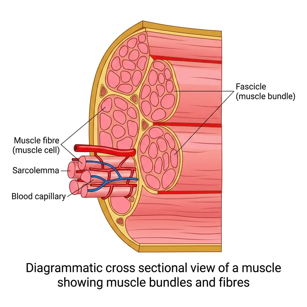
Structure of a Muscle: Muscle is composed of numerous muscle bundles (fascicles) wrapped in connective tissue and containing muscle cells and vascular capillaries.
🔬 Detailed Anatomical Description
Skeletal muscle consists of numerous muscle bundles called fascicles. Each fascicle is enclosed in a connective tissue sheath called epimysium/perimysium. Within each fascicle, there are many elongated muscle cells or muscle fibres surrounded by a plasma membrane called sarcolemma. Dense networks of blood capillaries infiltrate the tissue to supply oxygen and nutrients.
B MUSCLE FIBRE
| FEATURE | DETAIL |
|---|---|
| Muscle fibre membrane | Sarcolemma ⭐⭐⭐ |
| Cytoplasm | Sarcoplasm ⭐⭐⭐ |
| Nature of fibre | Syncytium / multinucleate ⭐⭐⭐ |
| Sarcoplasmic reticulum (SR) | Storehouse of Ca²⁺ ions ⭐⭐⭐ |
| Inside sarcoplasm | Many parallel filaments called myofilaments / myofibrils ⭐⭐⭐ |
C MYOFIBRIL BANDING PATTERN
| STRUCTURE | DETAIL |
|---|---|
| Myofibril appearance | Alternate dark and light bands ⭐⭐⭐ |
| Cause of striations | Distribution pattern of actin and myosin ⭐⭐⭐ |
NEET 2023: Striated appearance is due to distribution of actin and myosin.
NEET 2021: Myofibrils show alternate dark and light bands.
VI. BANDS, LINES AND SARCOMERE
| STRUCTURE | DETAIL |
|---|---|
| I-band / Isotropic / Light band | Contains actin (thin filament) ⭐⭐⭐ |
| A-band / Anisotropic / Dark band | Contains myosin (thick filament) ⭐⭐⭐ |
| Z-line | Elastic fibre present in centre of each I-band ⭐⭐⭐ |
| M-line | Thin fibrous membrane in middle of A-band ⭐⭐⭐ |
| Sarcomere | Portion of myofibril between two successive Z-lines; functional unit of muscle contraction ⭐⭐⭐ |
| H-zone | Central part of thick filament not overlapped by thin filament ⭐⭐⭐ |
Additional Notes ⭐⭐⭐
| FEATURE | DETAIL |
|---|---|
| Thin filaments | Firmly attached to Z-line ⭐⭐ |
| Thick filaments | Held together at centre by M-line ⭐⭐ |
| In resting state | Thin filaments partially overlap thick filaments ⭐⭐ |
| H-zone definition | Central gap between actin filaments extending through myosin in A-band ⭐⭐ |
NEET 2021: Z-line in centre of I-band; sarcomere = functional unit.
NEET 2013: H-zone = central part of thick filament not overlapped by actin.
Anatomy of a Muscle Fibre
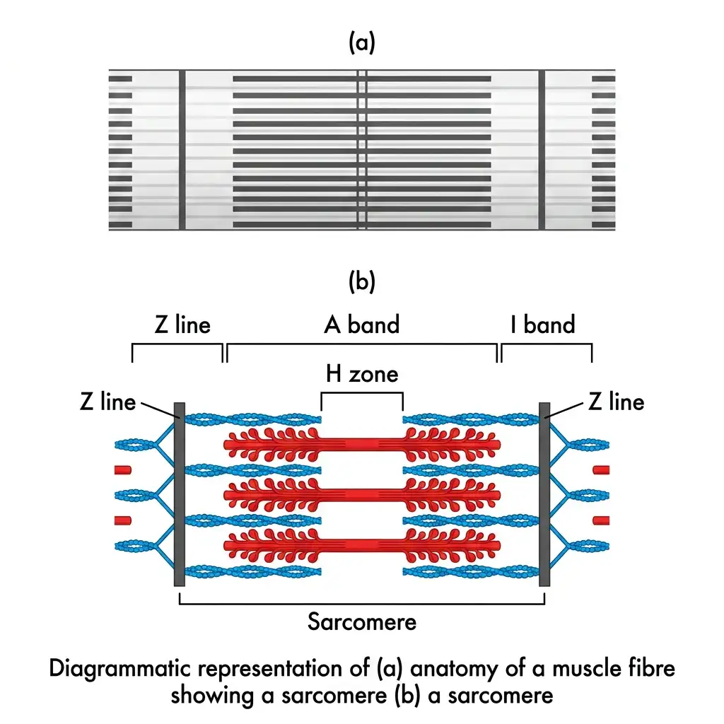
Anatomy of a Sarcomere: The functional unit of a myofibril, defined as the segment between two successive dark Z lines.
🔬 Detailed Anatomical Description
A muscle fibre contains myofibrils consisting of alternating light (I band) and dark (A band) bands. A sarcomere is the structural and functional unit of contraction located between two successive dark Z lines. The light I band contains thin actin filaments and is bisected by the Z line. The dark A band contains thick myosin filaments. The central region of thick filaments not overlapped by thin filaments is the H zone.
VII. STRUCTURE OF CONTRACTILE PROTEINS
A ACTIN (THIN FILAMENT)
| FEATURE | DETAIL |
|---|---|
| Thin filament made of | Two F-actins wound helically ⭐⭐⭐ |
| F-actin is | Polymer of G-actin monomers ⭐⭐⭐ |
| Tropomyosin | Two filamentous proteins run closely along F-actin ⭐⭐⭐ |
| Troponin | Complex protein found at regular intervals on tropomyosin ⭐⭐⭐ |
| Resting state | Troponin masks active binding sites for myosin on actin ⭐⭐⭐ |
B MYOSIN (THICK FILAMENT)
| FEATURE | DETAIL |
|---|---|
| Thick filament made of | Many monomeric proteins called meromyosins ⭐⭐⭐ |
| Meromyosin has two parts | Globular head with short arm (HMM) + Tail (LMM) ⭐⭐⭐ |
| Head projects outward as | Cross arm ⭐⭐ |
| Globular head function | Active ATPase enzyme ⭐⭐⭐ |
| Head has | ATP binding site and actin binding site ⭐⭐⭐ |
NEET 2021: Myosin head acts as ATPase.
Structure of Actin & Myosin
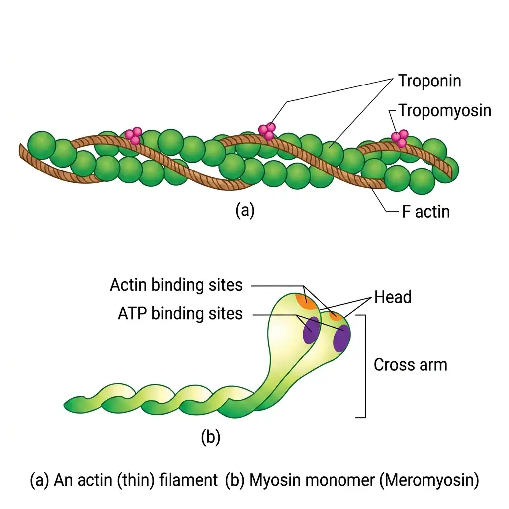
Contractile Proteins: Compares the multi-subunit thin actin filament with the monomeric units of the thick myosin filament (meromyosin).
🔬 Detailed Anatomical Description
Contractile proteins are composed of thin and thick filaments. (a) Thin filament is composed of two helical F-actin chains wrapped by tropomyosin. Troponin complexes are distributed regularly along tropomyosin to mask active binding sites in a relaxed state. (b) Thick filament consists of polymerized myosin. Each monomer (meromyosin) has a globular head (with ATP and actin binding sites) and a cross-arm.
VIII. MECHANISM OF MUSCLE CONTRACTION
A THEORY
| FEATURE | DETAIL |
|---|---|
| Best explained by | Sliding filament theory ⭐⭐⭐ |
| Principle | Thin filaments slide over thick filaments during contraction ⭐⭐⭐ |
NEET 2015, 2022: Muscle contraction follows sliding filament theory.
Sliding-Filament Theory
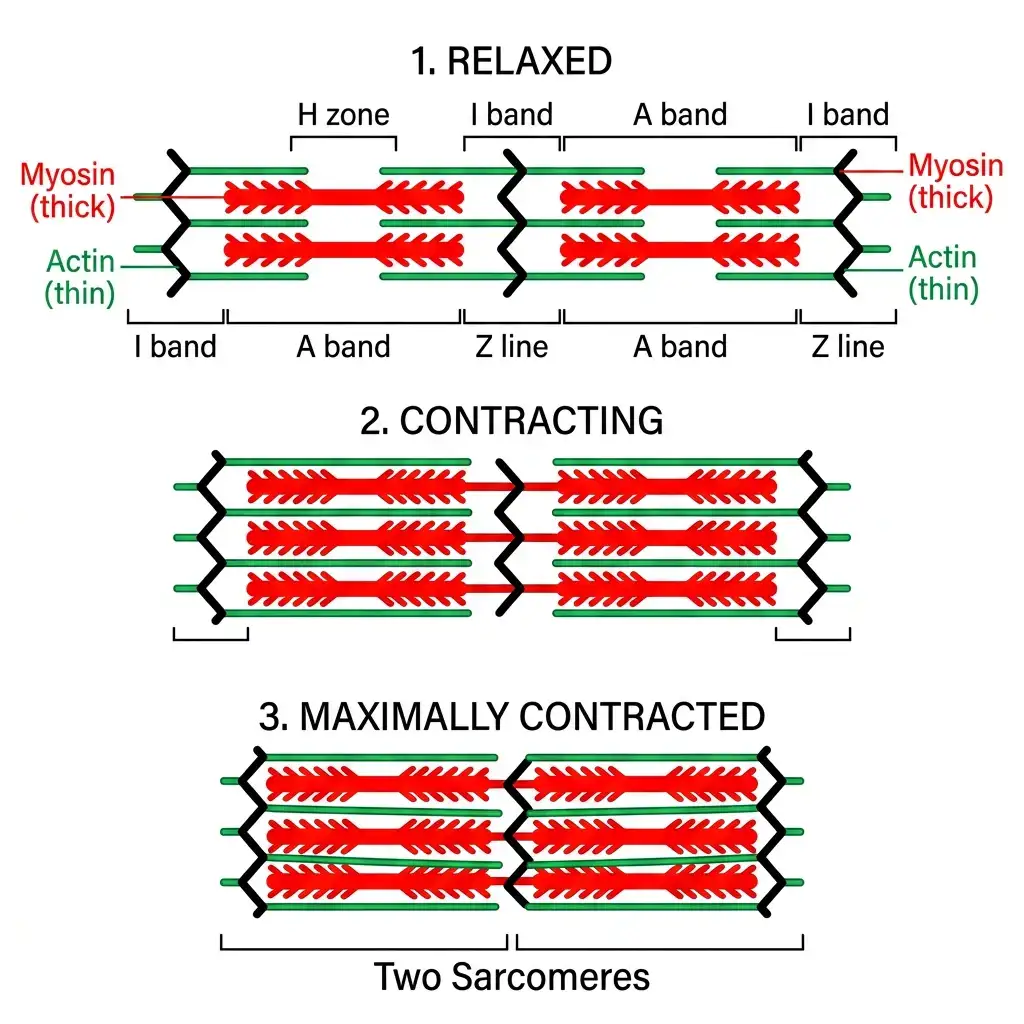
Sliding-Filament Model: Muscle contraction occurs when thin actin filaments slide past thick myosin filaments, shortening the sarcomere.
🔬 Detailed Anatomical Description
According to the sliding-filament theory, muscle contraction is achieved when thin filaments slide over thick filaments. During contraction, the sarcomere shortens because the Z lines are pulled closer. This leads to the progressive narrowing and eventual disappearance of the light I bands and H zones, while the width of the dark A bands remains completely unchanged.
B INITIATION OF CONTRACTION
| FEATURE | DETAIL |
|---|---|
| Signal sent by | CNS via motor neuron ⭐⭐⭐ |
| Motor unit | Motor neuron + muscle fibres connected to it ⭐⭐⭐ |
| Neuromuscular junction / Motor end plate | Junction between motor neuron and sarcolemma of muscle fibre ⭐⭐⭐ |
| Neurotransmitter released | Acetylcholine ⭐⭐ |
| Result | Generates action potential in sarcolemma ⭐⭐⭐ |
NEET 2014, 2024: Contraction initiated by motor neuron, not sensory neuron.
NEET 2014: Neuromuscular junction = junction of motor neuron and sarcolemma.
C ROLE OF CALCIUM
| FEATURE | DETAIL |
|---|---|
| Action potential causes | Release of Ca²⁺ from sarcoplasmic reticulum into sarcoplasm ⭐⭐⭐ |
| Ca²⁺ binds to | Troponin on actin filament ⭐⭐⭐ |
| Result | Removes masking of active sites on actin for myosin ⭐⭐⭐ |
NEET 2018, 2024: Ca²⁺ binds troponin → active sites on actin exposed.
D CROSS-BRIDGE FORMATION & SLIDING
| STEP | DETAIL |
|---|---|
| 1 | Myosin head hydrolyses ATP → ADP + Pi ⭐⭐⭐ |
| 2 | Using energy from ATP hydrolysis, myosin head binds exposed actin site → cross-bridge formed ⭐⭐⭐ |
| 3 | Myosin pulls actin toward centre of A-band ⭐⭐⭐ |
| 4 | Z-lines move inward → sarcomere shortens ⭐⭐⭐ |
| 5 | Myosin releases ADP and Pi, returns to relaxed stage ⭐⭐ |
| 6 | New ATP binds to myosin → cross-bridge breaks ⭐⭐⭐ |
| 7 | Cycle repeats if Ca²⁺ remains available ⭐⭐⭐ |
NEET 2024: Cross-bridge formation uses energy from ATP hydrolysis.
NEET 2021: Myosin head ATPase hydrolyses ATP → ADP + Pi.
Cross-Bridge Cycle Stages
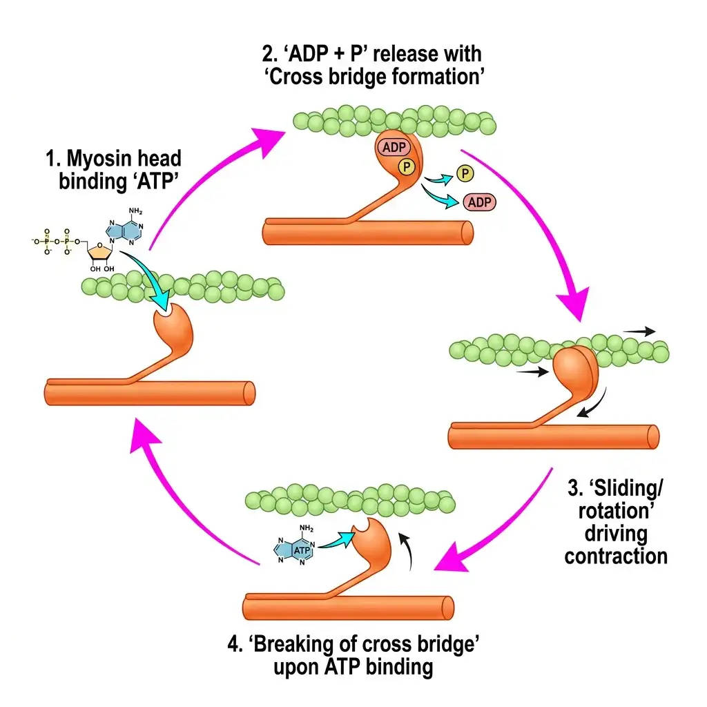
The Cross-Bridge Cycle: Illustrates active muscle contraction stages driven by ATP binding, hydrolysis, and sliding rotation.
🔬 Detailed Anatomical Description
Contraction begins when calcium ions bind to troponin, exposing actin binding sites. The myosin head hydrolyzes ATP to ADP+Pi and binds to actin, forming a cross-bridge. The head then rotates (power stroke), pulling the actin filament inward (sliding). ADP+Pi are released. A new ATP binds to the head, breaking the cross-bridge. The cycle repeats to continue muscle shortening.
E CHANGES DURING CONTRACTION
| STRUCTURE | CHANGE DURING CONTRACTION |
|---|---|
| A-band | Length remains constant ⭐⭐⭐ |
| I-band | Shortens / reduces ⭐⭐⭐ |
| H-zone | Reduces / disappears ⭐⭐⭐ |
| Z-lines | Pulled inward ⭐⭐⭐ |
| Sarcomere | Shortens ⭐⭐⭐ |
NEET 2021, 2022: A-band remains constant; I-band shortens.
NEET 2013, 2021: H-zone disappears.
F RELAXATION
| FEATURE | DETAIL |
|---|---|
| Relaxation occurs when | Ca²⁺ is pumped back into sarcoplasmic reticulum / sarcoplasmic cisternae ⭐⭐⭐ |
| Result | Actin sites become masked again; Z-lines return to original position ⭐⭐ |
G ADDITIONAL FACTS
| FEATURE | DETAIL |
|---|---|
| Reaction time of fibres | Can vary in different muscles ⭐ |
| Repeated activation of muscles | Causes lactic acid accumulation due to anaerobic breakdown of glycogen ⭐⭐⭐ |
| Result | Muscle fatigue ⭐⭐⭐ |
IX. RED VS WHITE MUSCLE FIBRES
| FEATURE | RED MUSCLE FIBRES | WHITE MUSCLE FIBRES |
|---|---|---|
| Myoglobin content | High ⭐⭐⭐ | Low ⭐⭐⭐ |
| Colour | Red ⭐⭐ | Pale / whitish ⭐⭐ |
| Mitochondria | More ⭐⭐⭐ | Fewer ⭐⭐⭐ |
| Sarcoplasmic reticulum | Less ⭐⭐ | More ⭐⭐ |
| Respiration type | Aerobic ⭐⭐⭐ | Anaerobic ⭐⭐⭐ |
X. SKELETAL SYSTEM — OVERVIEW
| FEATURE | DETAIL |
|---|---|
| Skeletal system consists of | Framework of bones and a few cartilages ⭐⭐⭐ |
| Bone and cartilage are | Specialised connective tissues ⭐⭐ |
| Bone matrix | Hard due to calcium salts ⭐⭐ |
| Cartilage matrix | Slightly pliable due to chondroitin salts ⭐⭐ |
| Total bones in human body | 206 bones ⭐⭐⭐ |
Main Divisions ⭐⭐⭐
| DIVISION | NO. OF BONES |
|---|---|
| Axial skeleton | 80 ⭐⭐⭐ |
| Appendicular skeleton | 126 ⭐⭐⭐ |
XI. AXIAL SKELETON
| COMPONENT | BONES |
|---|---|
| Skull | 22 (8 cranial + 14 facial) ⭐⭐⭐ |
| Ear ossicles | 6 (3 in each ear) ⭐⭐⭐ |
| Hyoid | 1 ⭐⭐⭐ |
| Vertebral column | 26 ⭐⭐⭐ |
| Sternum | 1 ⭐⭐⭐ |
| Ribs | 24 (12 pairs) ⭐⭐⭐ |
XII. SKULL
A SKULL BASICS
| FEATURE | DETAIL |
|---|---|
| Skull composed of | Cranial bones + Facial bones ⭐⭐⭐ |
| Cranial bones | 8 ⭐⭐⭐ |
| Facial bones | 14 ⭐⭐⭐ |
| Function of cranium | Protective outer covering for brain ⭐⭐ |
B CRANIAL BONES (STANDARD LIST)
| BONE | NUMBER |
|---|---|
| Frontal | 1 |
| Parietal | 2 |
| Temporal | 2 |
| Occipital | 1 |
| Sphenoid | 1 |
| Ethmoid | 1 |
C ASSOCIATED BONES
| STRUCTURE | DETAIL |
|---|---|
| Middle ear bones | Malleus, Incus, Stapes — 3 in each ear ⭐⭐⭐ |
| Hyoid | Single U-shaped bone at base of buccal cavity ⭐⭐⭐ |
NEET 2021: Hyoid = single U-shaped bone at base of buccal cavity.
D SKULL ARTICULATION
| FEATURE | DETAIL |
|---|---|
| Skull articulates with vertebral column by | Two occipital condyles ⭐⭐⭐ |
| Skull type | Dicondylic skull ⭐⭐⭐ |
NEET 2026 context: The occipital bone of the skull is articulated with the first vertebra.
Anatomy of Human Skull
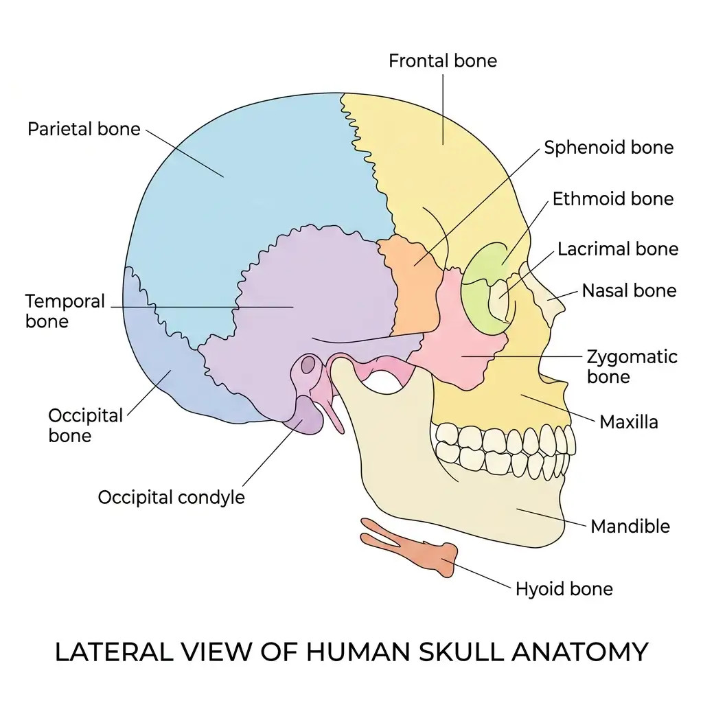
Structure of the Human Skull: Detailed lateral overview showing 22 bones divided into cranial and facial regions, plus the hyoid bone.
🔬 Detailed Anatomical Description
The human skull is composed of 22 bones grouped into: (1) Cranial bones (8 bones: frontal, parietal, temporal, occipital, sphenoid, ethmoid) forming a protective brain box; (2) Facial bones (14 bones: zygomatic, maxilla, mandible, nasal, lacrimal, palatine, inferior nasal conchae, vomer). The U-shaped hyoid bone sits at the base of the buccal cavity. The skull articulates via two occipital condyles (dicondylic skull).
XIII. VERTEBRAL COLUMN
| FEATURE | DETAIL |
|---|---|
| Number of vertebrae | 26 serially arranged bones ⭐⭐⭐ |
| Position | Dorsal side; extends from base of skull ⭐⭐ |
| Central hollow part of each vertebra | Neural canal ⭐⭐⭐ |
| Function of neural canal | Passage of spinal cord ⭐⭐⭐ |
Regions of Vertebral Column ⭐⭐⭐
| REGION | NUMBER |
|---|---|
| Cervical | 7 ⭐⭐⭐ |
| Thoracic | 12 ⭐⭐⭐ |
| Lumbar | 5 ⭐⭐⭐ |
| Sacral | 1 (fused) ⭐⭐⭐ |
| Coccygeal | 1 (fused) ⭐⭐⭐ |
Additional Facts ⭐⭐⭐
| FEATURE | DETAIL |
|---|---|
| First vertebra | Atlas ⭐⭐ |
| Cervical vertebrae in mammals | 7 in almost all mammals, including humans ⭐⭐⭐ |
| Cartilage between adjacent vertebrae | Present; allows limited movement ⭐⭐⭐ |
NEET 2022, 2026 (reinforced): Cartilage present between adjacent vertebrae. The joint between any two adjoining vertebrae is a cartilaginous joint.
NEET 2026 context: In humans, the number of cervical vertebrae is seven.
RE-NEET 2026: The correct order of arrangement of the vertebral column from the head to toe is: Cervical vertebra → Thoracic vertebra → Lumbar vertebra → Sacrum.
Vertebral Column Lateral View
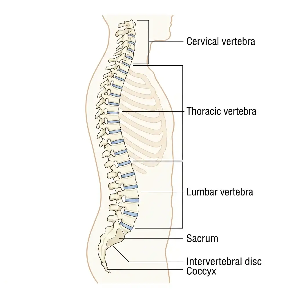
The Vertebral Column: A curved skeletal structure composed of 26 serially arranged vertebrae divided into cervical, thoracic, lumbar, sacral, and coccygeal zones.
🔬 Detailed Anatomical Description
The vertebral column consists of 26 serially arranged vertebrae: (1) Cervical (7 vertebrae, supporting the neck); (2) Thoracic (12 vertebrae, articulating with ribs); (3) Lumbar (5 vertebrae, supporting the lower back); (4) Sacrum (1 fused vertebra); (5) Coccyx (1 fused vertebra). Between each vertebral body lies an elastic intervertebral disc of fibrocartilage acting as a shock absorber.
XIV. STERNUM, RIBS AND RIB CAGE
A STERNUM
| FEATURE | DETAIL |
|---|---|
| Sternum | Flat bone on ventral midline of thorax ⭐⭐⭐ |
NEET 2021: Sternum is a flat bone.
B RIBS
| FEATURE | DETAIL |
|---|---|
| Total ribs | 12 pairs ⭐⭐⭐ |
| Nature | Thin, flat bones ⭐⭐ |
| Dorsal attachment | Vertebral column ⭐⭐⭐ |
| Ventral attachment | Sternum (directly or indirectly) ⭐⭐⭐ |
| Dorsal end | Has two articulation surfaces → ribs are bicephalic ⭐⭐⭐ |
NEET 2020, 2026 (reinforced): All ribs except the last 2 pairs are bicephalic.
C TYPES OF RIBS
| RIB PAIRS | NAME | ANOTHER NAME | DETAIL |
|---|---|---|---|
| 1st–7th | Vertebrosternal ribs | True ribs ⭐⭐⭐ | Directly connected to sternum by hyaline cartilage |
| 8th–10th | Vertebrochondral ribs | False ribs ⭐⭐⭐ | Indirectly connected to sternum through 7th rib |
| 11th–12th | Vertebral ribs | Floating ribs ⭐⭐⭐ | No ventral connection to sternum |
D RIB CAGE
| FEATURE | DETAIL |
|---|---|
| Rib cage formed by | Thoracic vertebrae + ribs + sternum ⭐⭐⭐ |
NEET 2017, 2019: True / false / floating rib classification.
NEET 2020: Floating ribs = 11th and 12th; ribs are bicephalic.
Structure of Rib Cage
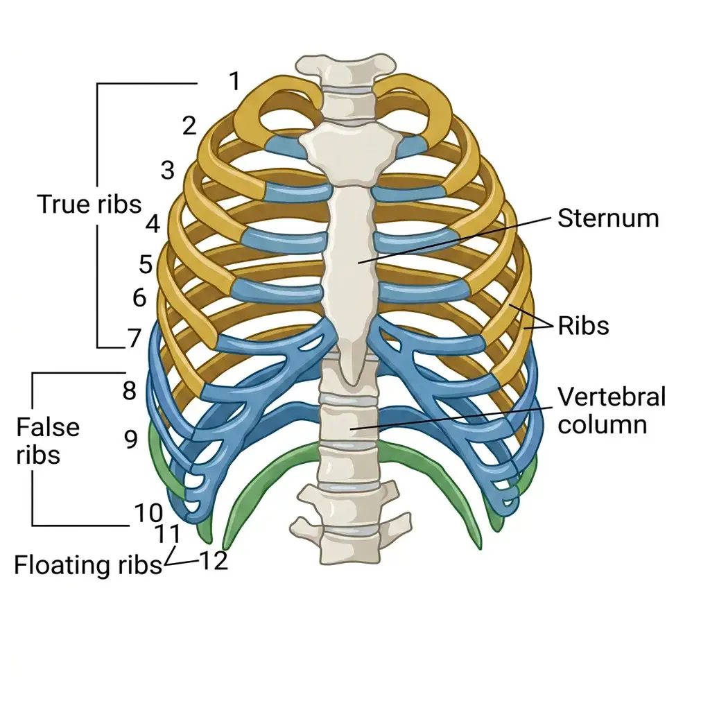
The Rib Cage: Protective skeletal cage composed of 12 pairs of ribs (true, false, and floating) connected to the sternum and thoracic vertebrae.
🔬 Detailed Anatomical Description
The rib cage protects the thoracic organs. It consists of: (1) True ribs (pairs 1 to 7, connected directly to sternum via hyaline cartilage); (2) False ribs (pairs 8, 9, 10, connected to the seventh rib); (3) Floating ribs (pairs 11 and 12, not attached anteriorly). Ribs are dorsally connected to the thoracic vertebrae and ventrally to the flat sternum breastbone.
XV. APPENDICULAR SKELETON
| COMPONENT | BONES |
|---|---|
| Pectoral girdles | 4 (2 scapulae + 2 clavicles) ⭐⭐⭐ |
| Forelimbs | 60 ⭐⭐⭐ |
| Pelvic girdle | 2 (2 coxal bones) ⭐⭐⭐ |
| Hind limbs | 60 ⭐⭐⭐ |
A LIMB BONE COUNTS
Forelimb (each limb = 30 bones) ⭐⭐⭐
| BONE | NUMBER |
|---|---|
| Humerus | 1 |
| Radius | 1 |
| Ulna | 1 |
| Carpals (wrist bones) | 8 |
| Metacarpals (palm bones) | 5 |
| Phalanges (digits) | 14 |
Pectoral Girdle & Upper Arm
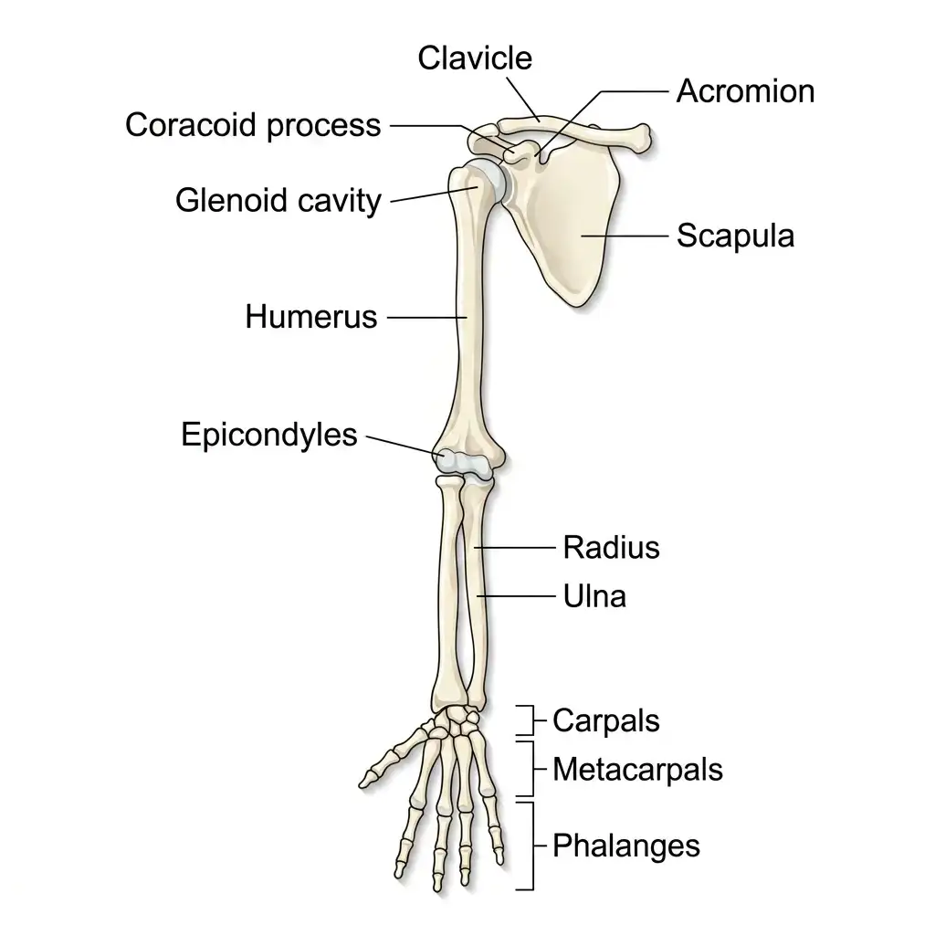
The Pectoral Girdle & Forelimb: Shows shoulder joints and arm bones (humerus, radius, ulna, hand) enabling flexible upper body mobility.
🔬 Detailed Anatomical Description
The pectoral girdle connects the forelimbs to the axial skeleton, comprising the clavicle (collarbone) and scapula (shoulder blade with glenoid cavity). The right upper arm features a single long humerus bone articulating with the scapula. The forearm is composed of parallel radius and ulna bones, which articulate with 8 carpals (wrist), 5 metacarpals (palm), and 14 phalanges (fingers).
Hind limb (each limb = 30 bones) ⭐⭐⭐
| BONE | NUMBER |
|---|---|
| Femur | 1 |
| Tibia | 1 |
| Fibula | 1 |
| Tarsals (ankle bones) | 7 |
| Metatarsals | 5 |
| Phalanges | 14 |
Pelvic Girdle & Lower Limb
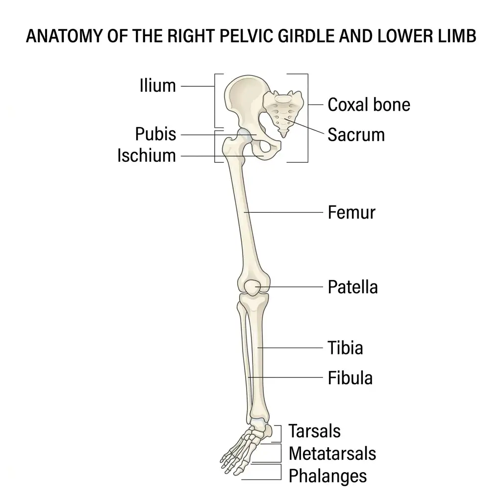
The Pelvic Girdle & Hindlimb: Shows the weight-bearing hip joint (ilium, pubis, ischium) and leg bones (femur, patella, tibia, fibula, foot).
🔬 Detailed Anatomical Description
The pelvic girdle consists of two coxal bones, each formed by the fusion of ilium, pubis, and ischium. They meet anteriorly at the pubic symphysis. The hindlimb is composed of: the femur (thigh bone, longest bone in the body); the patella (kneecap); the tibia (shinbone) and fibula; 7 tarsals (ankle bones); 5 metatarsals (instep); and 14 phalanges (toes).
Additional Facts ⭐⭐⭐
| FEATURE | DETAIL |
|---|---|
| Longest bone in body | Femur ⭐⭐⭐ |
| Patella | Cup-shaped bone covering knee ventrally; knee cap ⭐⭐⭐ |
XVI. PECTORAL GIRDLE
| FEATURE | DETAIL |
|---|---|
| Each half consists of | Scapula + Clavicle ⭐⭐⭐ |
| Clavicle | Commonly called collar bone ⭐⭐ |
Scapula ⭐⭐⭐
| FEATURE | DETAIL |
|---|---|
| Nature | Large, triangular, flat bone ⭐⭐⭐ |
| Position | Dorsal part of thorax between 2nd and 7th ribs ⭐⭐⭐ |
| Elevated ridge on scapula | Spine ⭐⭐ |
| Expanded process of spine | Acromion ⭐⭐⭐ |
| Acromion articulates with | Clavicle ⭐⭐⭐ |
| Depression below acromion | Glenoid cavity ⭐⭐⭐ |
| Glenoid cavity articulates with | Head of humerus to form shoulder joint ⭐⭐⭐ |
NEET 2020, 2021: Scapula lies between 2nd and 7th ribs.
NEET 2020: Acromion articulates with clavicle.
NEET 2015: Glenoid cavity articulates with humerus.
XVII. PELVIC GIRDLE
| FEATURE | DETAIL |
|---|---|
| Pelvic girdle consists of | Two coxal bones ⭐⭐⭐ |
| Each coxal bone formed by fusion of | Ilium, Ischium, Pubis ⭐⭐⭐ |
| Acetabulum | Cavity at fusion point of these bones ⭐⭐⭐ |
| Acetabulum articulates with | Thigh bone (femur) ⭐⭐⭐ |
| Two halves meet ventrally to form | Pubic symphysis ⭐⭐⭐ |
| Pubic symphysis contains | Fibrous cartilage ⭐⭐⭐ |
NEET 2023: Pubic symphysis contains fibrous cartilage.
XVIII. JOINTS
A BASIC CONCEPT
| FEATURE | DETAIL |
|---|---|
| Joint | Point of contact between bones or between bone and cartilage ⭐⭐⭐ |
| Importance | Essential for movement; joint acts as fulcrum ⭐⭐ |
| Types | Fibrous, Cartilaginous, Synovial ⭐⭐⭐ |
B TYPES OF JOINTS
| JOINT TYPE | MOVEMENT | EXAMPLE |
|---|---|---|
| Fibrous joint | No movement ⭐⭐⭐ | Skull bones / cranial sutures ⭐⭐⭐ |
| Cartilaginous joint | Limited movement ⭐⭐⭐ | Between adjacent vertebrae ⭐⭐⭐ |
| Synovial joint | Considerable movement ⭐⭐⭐ | Shoulder, knee, thumb etc. ⭐⭐⭐ |
C SYNOVIAL JOINTS — IMPORTANT TYPES
| SYNOVIAL JOINT | EXAMPLE |
|---|---|
| Ball and socket joint | Between humerus and pectoral girdle; also hip (femur-acetabulum) ⭐⭐⭐ |
| Hinge joint | Knee, elbow, phalanges ⭐⭐⭐ |
| Pivot joint | Between atlas and axis ⭐⭐⭐ |
| Gliding joint | Between carpals ⭐⭐⭐ |
| Saddle joint | Between carpal and metacarpal of thumb ⭐⭐⭐ |
D SYNOVIAL JOINT FEATURE
| FEATURE | DETAIL |
|---|---|
| Synovial joints have | Fluid-filled synovial cavity ⭐⭐⭐ |
| Synovial fluid function | Allows smooth movement ⭐⭐ |
NEET 2015, 2017, 2021, 2024: Fibrous joints = skull sutures.
NEET 2013, 2017, 2023, 2024: Cartilaginous joint = between adjacent vertebrae.
NEET 2016, 2023, 2024: Ball and socket = shoulder joint.
NEET 2016, 2024: Hinge = knee / elbow / phalanges.
NEET 2016, 2017, 2024: Pivot = atlas-axis.
NEET 2014, 2016, 2024: Gliding = between carpals.
NEET 2018: Saddle = thumb joint.
XIX. DISORDERS OF MUSCULAR AND SKELETAL SYSTEM
| DISORDER | KEY FEATURES |
|---|---|
| Myasthenia gravis | Autoimmune disorder affecting neuromuscular junction; causes fatigue, weakening and paralysis of skeletal muscles ⭐⭐⭐ |
| Muscular dystrophy | Genetic / inherited disorder; progressive degeneration of skeletal muscles ⭐⭐⭐ |
| Tetany | Rapid spasms / wild contractions due to low Ca²⁺ in body fluids ⭐⭐⭐ |
| Arthritis | Inflammation of joints ⭐⭐ |
| Osteoporosis | Age-related disorder; decreased bone mass and increased fracture risk; common cause = decreased estrogen ⭐⭐⭐ |
| Gout | Inflammation of joints due to uric acid crystal accumulation ⭐⭐⭐ |
Additional PYQ-linked Autoimmune Facts ⭐⭐
| DISORDER | DETAIL |
|---|---|
| Rheumatoid arthritis | Autoimmune disorder ⭐⭐ |
| Systemic Lupus Erythematosus (SLE) | Autoimmune disorder ⭐⭐ |
Important Trap ⭐⭐
| TERM | MEANING |
|---|---|
| Tetany | Spasm due to low Ca²⁺ ⭐⭐⭐ |
| Tetanus | Sustained contraction / lack of relaxation between successive stimuli ⭐⭐ |
NEET 2022, 2024: Myasthenia gravis = autoimmune NMJ disorder.
NEET 2012, 2013, 2021, 2022, 2024: Muscular dystrophy = genetic skeletal muscle degeneration.
NEET 2016, 2022: Tetany due to low Ca²⁺.
NEET 2012, 2013, 2016, 2022: Osteoporosis due to decreased bone mass; estrogen decline common cause.
NEET 2022, 2024: Gout = uric acid crystals in joints.
XX. RAPID REVISION — KEY COMPARISON TABLES
TABLE 1: Types of Movement ⭐⭐⭐
| TYPE | EXAMPLES | MECHANISM |
|---|---|---|
| Amoeboid | Macrophages, WBCs, Amoeba | Pseudopodia + microfilaments |
| Ciliary | Trachea, female reproductive tract | Coordinated cilia beating |
| Muscular | Limbs, jaws, tongue | Muscle contraction |
| Flagellar | Spermatozoa, sponges, Euglena | Flagella |
TABLE 2: Types of Muscles ⭐⭐⭐
| FEATURE | SKELETAL | VISCERAL | CARDIAC |
|---|---|---|---|
| Appearance | Striated | Smooth / unstriated | Striated |
| Control | Voluntary | Involuntary | Involuntary |
| Location | Attached to skeleton | Wall of hollow organs | Heart |
| Main role | Locomotion / posture | Movement of contents | Pumping blood |
TABLE 3: Bands and Lines of Sarcomere ⭐⭐⭐
| STRUCTURE | LOCATION / PROTEIN |
|---|---|
| I-band | Light band; actin |
| A-band | Dark band; myosin |
| Z-line | Centre of I-band |
| M-line | Centre of A-band |
| H-zone | Central part of thick filament not overlapped by thin filament |
| Sarcomere | Between two successive Z-lines |
TABLE 4: Contractile Proteins ⭐⭐⭐
| PROTEIN | STRUCTURE / FEATURE |
|---|---|
| Actin | Thin filament; 2 F-actins made of G-actin monomers |
| Tropomyosin | Runs along actin |
| Troponin | On tropomyosin; masks actin active sites in resting state |
| Myosin | Thick filament; made of meromyosins |
| HMM | Head + short arm |
| LMM | Tail |
| Myosin head | ATPase; ATP-binding + actin-binding site |
TABLE 5: Changes During Contraction ⭐⭐⭐
| STRUCTURE | CHANGE |
|---|---|
| A-band | No change |
| I-band | Shortens |
| H-zone | Disappears / shortens |
| Z-line | Pulled inward |
| Sarcomere | Shortens |
TABLE 6: Red vs White Fibres ⭐⭐⭐
| FEATURE | RED FIBRES | WHITE FIBRES |
|---|---|---|
| Myoglobin | High | Low |
| Mitochondria | More | Fewer |
| SR | Less | More |
| Respiration | Aerobic | Anaerobic |
TABLE 7: Axial vs Appendicular Skeleton ⭐⭐⭐
| DIVISION | BONES |
|---|---|
| Axial | 80 |
| Appendicular | 126 |
| Total | 206 |
TABLE 8: Axial Skeleton Breakdown ⭐⭐⭐
| COMPONENT | NUMBER |
|---|---|
| Cranial bones | 8 |
| Facial bones | 14 |
| Ear ossicles | 6 |
| Hyoid | 1 |
| Vertebral column | 26 |
| Sternum | 1 |
| Ribs | 24 |
TABLE 9: Vertebral Column ⭐⭐⭐
| REGION | NUMBER |
|---|---|
| Cervical | 7 |
| Thoracic | 12 |
| Lumbar | 5 |
| Sacral | 1 (fused) |
| Coccygeal | 1 (fused) |
TABLE 10: Ribs Classification ⭐⭐⭐
| RIB PAIR | TYPE | ALSO CALLED |
|---|---|---|
| 1–7 | Vertebrosternal | True ribs |
| 8–10 | Vertebrochondral | False ribs |
| 11–12 | Vertebral | Floating ribs |
TABLE 11: Limb Bone Counts ⭐⭐⭐
| BONE TYPE | FORELIMB | HIND LIMB |
|---|---|---|
| Long bones | Humerus, Radius, Ulna | Femur, Tibia, Fibula |
| Wrist / Ankle bones | Carpals (8) | Tarsals (7) |
| Palm / Sole bones | Metacarpals (5) | Metatarsals (5) |
| Digits | Phalanges (14) | Phalanges (14) |
TABLE 12: Types of Joints ⭐⭐⭐
| JOINT TYPE | MOVEMENT | EXAMPLE |
|---|---|---|
| Fibrous | No movement | Skull sutures |
| Cartilaginous | Limited movement | Between adjacent vertebrae |
| Synovial | Free movement | Shoulder, knee, thumb |
TABLE 13: Synovial Joint Examples ⭐⭐⭐
| JOINT | EXAMPLE |
|---|---|
| Ball and socket | Shoulder / Hip |
| Hinge | Knee / Elbow / Phalanges |
| Pivot | Atlas–Axis |
| Gliding | Carpals |
| Saddle | Thumb |
TABLE 14: Disorders ⭐⭐⭐
| DISORDER | KEY POINT |
|---|---|
| Myasthenia gravis | Autoimmune; neuromuscular junction |
| Muscular dystrophy | Genetic degeneration of skeletal muscle |
| Tetany | Low Ca²⁺ spasms |
| Arthritis | Joint inflammation |
| Osteoporosis | Age-related bone mass loss |
| Gout | Uric acid crystal deposition in joints |
XXI. KEY NUMBERS — QUICK REFERENCE
| PARAMETER | VALUE |
|---|---|
| Muscle contribution to body weight | 40–50% |
| Total bones in skeleton | 206 |
| Axial skeleton | 80 bones |
| Appendicular skeleton | 126 bones |
| Cranial bones | 8 |
| Facial bones | 14 |
| Ear ossicles | 6 |
| Hyoid | 1 |
| Vertebrae | 26 |
| Cervical vertebrae | 7 |
| Thoracic vertebrae | 12 |
| Lumbar vertebrae | 5 |
| Ribs | 12 pairs |
| True ribs | 1st–7th |
| False ribs | 8th–10th |
| Floating ribs | 11th–12th |
| Bones in each limb | 30 |
| Carpals | 8 |
| Metacarpals | 5 |
| Phalanges per limb | 14 |
| Tarsals | 7 |
| Myosin head | ATPase |
| Functional unit of contraction | Sarcomere |
XXII. COMMON EXAM TRAPS — QUICK REFERENCE
| TRAP / QUESTION | CORRECT ANSWER |
|---|---|
| All movements are locomotion? | No |
| All locomotions are movements? | Yes |
| Amoeboid movement in human body shown by? | Macrophages and leucocytes |
| Ciliary movement helps passage of ova where? | Female reproductive tract |
| Flagellar movement example in humans? | Spermatozoa |
| Muscles are ectodermal? | No, mesodermal |
| Striped voluntary muscle? | Skeletal muscle |
| Unstriped involuntary muscle? | Visceral / smooth muscle |
| Cardiac muscle voluntary? | No, involuntary |
| Fascicles held by? | Fascia |
| Muscle fibre is uninucleate? | No, multinucleate syncytium |
| Storehouse of Ca²⁺ in muscle? | Sarcoplasmic reticulum |
| I-band contains? | Actin |
| A-band contains? | Myosin |
| Z-line location? | Centre of I-band |
| M-line location? | Centre of A-band |
| Functional unit of muscle contraction? | Sarcomere |
| H-zone contains both actin and myosin? | No — only thick filament central part not overlapped |
| Thin filament made of? | Actin + tropomyosin + troponin |
| Myosin head enzyme? | ATPase |
| Contraction explained by? | Sliding filament theory |
| Thin filaments slide over? | Thick filaments |
| Signal carried by sensory neuron? | No, motor neuron |
| NMJ = ? | Junction of motor neuron and sarcolemma |
| Neurotransmitter at NMJ? | Acetylcholine |
| Ca²⁺ binds myosin? | No, binds troponin |
| Ca²⁺ source in muscle? | Sarcoplasmic reticulum |
| During contraction A-band changes? | No change |
| During contraction I-band? | Shortens |
| H-zone during contraction? | Disappears / reduces |
| Z-lines move? | Inward |
| Cause of muscle fatigue? | Lactic acid accumulation |
| Red fibres are anaerobic? | No, aerobic |
| White fibres have more mitochondria? | No, fewer |
| Total bones in human skeleton? | 206 |
| Axial skeleton bones? | 80 |
| Appendicular skeleton bones? | 126 |
| Skull articulates with vertebral column by? | Two occipital condyles |
| Cervical vertebrae in mammals? | 7 |
| Sternum type of bone? | Flat bone |
| Ribs are monocephalic? | No, bicephalic |
| Floating ribs are 10th and 11th? | No, 11th and 12th |
| Scapula location? | Between 2nd and 7th ribs |
| Acromion articulates with? | Clavicle |
| Glenoid cavity articulates with? | Humerus |
| Acetabulum articulates with? | Femur |
| Pubic symphysis contains? | Fibrous cartilage |
| Fibrous joints allow movement? | No |
| Cartilaginous joints allow? | Limited movement |
| Synovial joints have? | Synovial cavity with synovial fluid |
| Pivot joint example? | Atlas-axis |
| Saddle joint example? | Thumb |
| Myasthenia gravis is genetic? | No, autoimmune NMJ disorder |
| Muscular dystrophy autoimmune? | No, genetic disorder |
| Tetany due to low Ca²⁺? | Yes |
| Osteoporosis due to progesterone deficiency? | No, commonly due to decreased estrogen |
| Gout due to uric acid crystals? | Yes |
| Tetany = tetanus? | No |