I. INTRODUCTION & COORDINATION
| FEATURE | DETAIL |
|---|---|
| Coordination | Process through which two or more organs interact and complement each other’s functions ⭐⭐ |
| Main coordinating systems in human body | Neural system + Endocrine system ⭐⭐⭐ |
| Neural system role | Provides an organised network of point-to-point connections for quick coordination ⭐⭐⭐ |
| Endocrine system role | Provides chemical integration through hormones ⭐⭐⭐ |
II. NEURAL SYSTEM IN ANIMALS
| ORGANISM LEVEL | NEURAL ORGANISATION |
|---|---|
| Lower invertebrates (e.g. Hydra) | Simple network of neurons ⭐ |
| Insects | Brain + ganglia + neural tissues ⭐ |
| Vertebrates | More developed neural system ⭐⭐ |
III. HUMAN NEURAL SYSTEM
A TWO MAIN DIVISIONS
| DIVISION | FULL FORM | COMPONENTS / ROLE |
|---|---|---|
| CNS | Central Neural System | Brain + spinal cord; site of information processing and control ⭐⭐⭐ |
| PNS | Peripheral Neural System | All nerves associated with CNS ⭐⭐⭐ |
Basic fact: CNS = brain + spinal cord; PNS = nerves associated with CNS.
IV. PERIPHERAL NERVOUS SYSTEM (PNS)
A TYPES OF NERVE FIBRES
| TYPE | DIRECTION OF IMPULSE |
|---|---|
| Afferent fibres | Carry impulses from tissues / organs → CNS ⭐⭐⭐ |
| Efferent fibres | Carry impulses from CNS → peripheral tissues / organs ⭐⭐⭐ |
B DIVISIONS OF PNS
| DIVISION | FUNCTION |
|---|---|
| Somatic neural system | Relays impulses from CNS → skeletal muscles ⭐⭐⭐ |
| Autonomic neural system (ANS) | Transmits impulses from CNS → involuntary organs and smooth muscles ⭐⭐⭐ |
C AUTONOMIC NERVOUS SYSTEM
| PART | GENERAL ROLE |
|---|---|
| Sympathetic neural system | Part of ANS ⭐⭐ |
| Parasympathetic neural system | Part of ANS ⭐⭐ |
D VISCERAL NERVOUS SYSTEM
| FEATURE | DETAIL |
|---|---|
| Visceral nervous system | Part of PNS in which impulses travel from CNS to viscera and from viscera to CNS ⭐⭐ |
V. NEURON — STRUCTURAL AND FUNCTIONAL UNIT
| FEATURE | DETAIL |
|---|---|
| Neuron | Structural and functional unit of neural system ⭐⭐⭐ |
| Nature | Highly specialised cell that can detect, receive and transmit stimuli ⭐⭐ |
A THREE MAIN PARTS OF A NEURON
| PART | DETAIL |
|---|---|
| Cell body / Cyton | Contains cytoplasm, cell organelles and Nissl’s granules ⭐⭐⭐ |
| Dendrites | Short, branched fibres; also contain Nissl’s granules; carry impulse towards cell body ⭐⭐⭐ |
| Axon | Long fibre; transmits impulse away from cell body ⭐⭐⭐ |
B AXON TERMINAL
| FEATURE | DETAIL |
|---|---|
| Distal end of axon | Branched ⭐⭐ |
| Terminal swelling | Synaptic knob ⭐⭐⭐ |
| Synaptic knob contains | Synaptic vesicles filled with neurotransmitters ⭐⭐⭐ |
NEET 2013, 2024: Synaptic knob = bulb-like axon terminal containing synaptic vesicles.
Structure of a Neuron
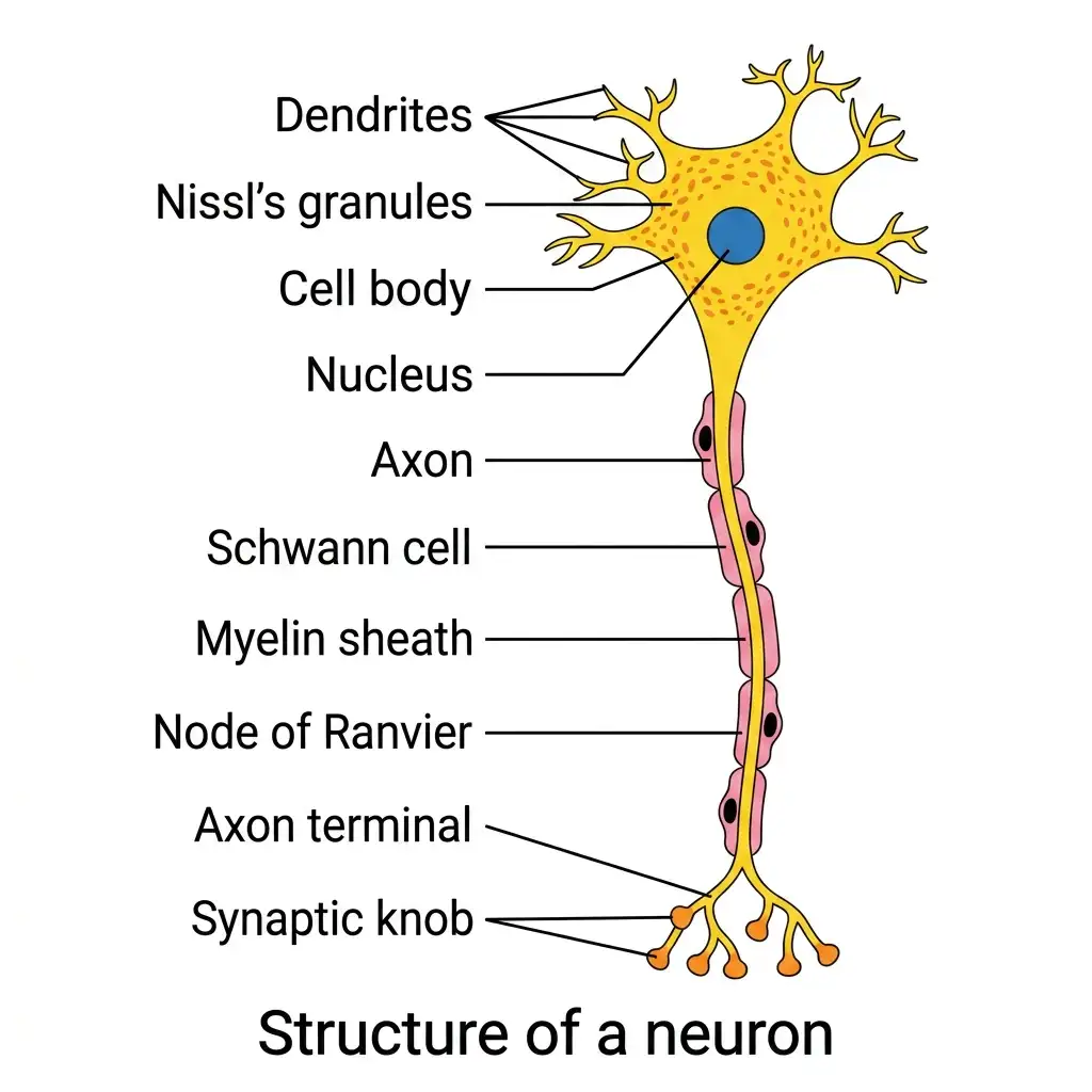
Structure of a Neuron: The structural and functional unit of the neural system, comprising dendrites, cell body, axon, and synaptic terminals.
🔬 Detailed Anatomical Description
A neuron is a highly specialized cell composed of three main parts: (1) Cell body (soma) containing cytoplasm, typical organelles, and granular bodies called Nissl's granules; (2) Dendrites, highly branched short fibers projecting out of the cell body to receive incoming signals; (3) Axon, a long single fiber conducting impulses away to synaptic knobs. Myelinated neurons are wrapped in a lipid-rich myelin sheath formed by Schwann cells, separated by nodes of Ranvier.
VI. NISSL BODIES / NISSL’S GRANULES
| FEATURE | DETAIL |
|---|---|
| Present in | Cell body and dendrites ⭐⭐⭐ |
| Absent in | Axon ⭐⭐ |
| Composition | Mainly free ribosomes and rough endoplasmic reticulum (RER) ⭐⭐ |
NEET 2018: Nissl bodies composed mainly of RER + ribosomes.
VII. TYPES OF NEURONS
| TYPE | STRUCTURE | EXAMPLE |
|---|---|---|
| Multipolar neuron | 1 axon + 2 or more dendrites ⭐⭐⭐ | Cerebral cortex ⭐⭐⭐ |
| Bipolar neuron | 1 axon + 1 dendrite ⭐⭐⭐ | Retina of eye ⭐⭐⭐ |
| Unipolar neuron | Cell body with one axon only ⭐⭐⭐ | Usually found in embryonic stage ⭐⭐⭐ |
NEET 2022, 2024, 2026 (reinforced): Multipolar neurons are found in the cerebral cortex; bipolar neurons are found in the retina.
VIII. MYELINATED VS NON-MYELINATED NERVE FIBRES
| FEATURE | MYELINATED NERVE FIBRE | NON-MYELINATED NERVE FIBRE |
|---|---|---|
| Covering | Enveloped by Schwann cells forming myelin sheath ⭐⭐⭐ | Enclosed by Schwann cell but no myelin sheath formed ⭐⭐⭐ |
| Location | Found in spinal and cranial nerves ⭐⭐⭐ | Commonly found in autonomic and somatic neural systems ⭐⭐ |
| Node of Ranvier | Present ⭐⭐⭐ | Absent ⭐⭐ |
Additional High-Yield Fact ⭐⭐
| CELL PRODUCING MYELIN | LOCATION |
|---|---|
| Schwann cells | PNS ⭐⭐ |
| Oligodendrocytes | CNS ⭐⭐ |
NEET 2017, 2024, 2026 (reinforced): Myelin sheath in the PNS is formed by Schwann cells; in the CNS it is formed by oligodendrocytes.
Node of Ranvier ⭐⭐⭐
| FEATURE | DETAIL |
|---|---|
| Definition | Gap between two adjacent myelin sheaths ⭐⭐⭐ |
IX. GENERATION AND CONDUCTION OF NERVE IMPULSE
A RESTING MEMBRANE / POLARISATION
| FEATURE | DETAIL |
|---|---|
| At rest, axonal membrane is more permeable to | K⁺ ions ⭐⭐⭐ |
| At rest, axonal membrane is nearly impermeable to | Na⁺ ions ⭐⭐⭐ |
| Membrane also impermeable to | Negatively charged proteins in axoplasm ⭐⭐⭐ |
Ionic Distribution at Rest ⭐⭐⭐
| SITE | MAJOR CONTENTS |
|---|---|
| Inside axon (axoplasm) | High K⁺, high negatively charged proteins, low Na⁺ ⭐⭐⭐ |
| Outside axon | High Na⁺, low K⁺ ⭐⭐⭐ |
Sodium-Potassium Pump ⭐⭐⭐
| FEATURE | DETAIL |
|---|---|
| Function | Maintains ionic gradient across resting membrane ⭐⭐⭐ |
| Transport | 3 Na⁺ out and 2 K⁺ in ⭐⭐⭐ |
Result ⭐⭐⭐
| SURFACE | CHARGE |
|---|---|
| Outer surface of membrane | Positive ⭐⭐⭐ |
| Inner surface of membrane | Negative ⭐⭐⭐ |
| TERM | MEANING |
|---|---|
| Polarised membrane | Outer surface +ve, inner surface –ve ⭐⭐⭐ |
| Resting potential | Electrical potential difference across resting membrane ⭐⭐⭐ |
NEET 2011: Resting membrane more permeable to K⁺ than Na⁺.
NEET 2013: Inside axon = high K⁺ and proteins, low Na⁺.
B DEPOLARISATION / ACTION POTENTIAL
| FEATURE | DETAIL |
|---|---|
| When stimulus is applied | Membrane at stimulated site becomes freely permeable to Na⁺ ⭐⭐⭐ |
| Result | Rapid influx of Na⁺ ⭐⭐⭐ |
| Polarity reverses at stimulus site | Outer surface becomes negative, inner surface becomes positive ⭐⭐⭐ |
| This state is called | Depolarisation ⭐⭐⭐ |
| Electrical potential difference at stimulated site | Action potential / Nerve impulse ⭐⭐⭐ |
Current Flow During Impulse ⭐⭐⭐
| SURFACE | DIRECTION OF CURRENT |
|---|---|
| Outer surface | From B to A ⭐⭐⭐ |
| Inner surface | From A to B ⭐⭐⭐ |
This causes the adjacent membrane to depolarise and the impulse moves forward.
C REPOLARISATION
| FEATURE | DETAIL |
|---|---|
| Rise in Na⁺ permeability | Very short-lived ⭐⭐ |
| Then membrane becomes more permeable to | K⁺ ⭐⭐⭐ |
| K⁺ diffuses | Outside the membrane ⭐⭐ |
| Result | Resting potential is restored = repolarisation ⭐⭐⭐ |
Impulse Conduction along Axon
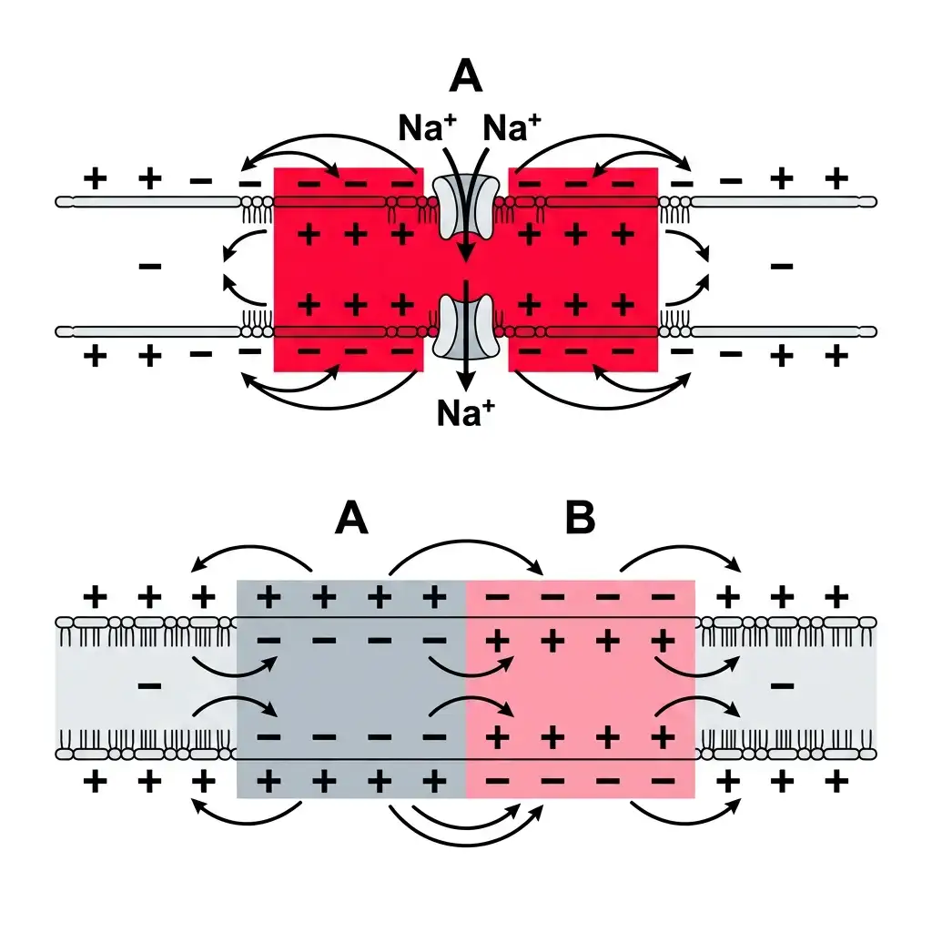
Nerve Impulse Conduction: Details polarization, depolarization (action potential), and propagation across axonal membranes.
🔬 Detailed Anatomical Description
Neurons are excitable cells due to polarized axonal membranes. (1) Resting State: High outer Na+ concentration and high inner K+ concentration create a negative resting potential inside (-70 mV). (2) Depolarization (Point A): A stimulus opens Na+ channels, causing rapid influx and reversing polarity (+30 mV, creating an action potential). (3) Propagation (Point B): Local currents flow between A and B, propagating the action potential forward, while Point A undergoes repolarization.
X. SYNAPSE & TRANSMISSION OF IMPULSES
| FEATURE | DETAIL |
|---|---|
| Synapse | Junction through which impulse passes from one neuron to another ⭐⭐⭐ |
| Formed by | Pre-synaptic neuron and post-synaptic neuron ⭐⭐⭐ |
| Synaptic cleft | Gap between them; may or may not be present ⭐⭐ |
XI. TYPES OF SYNAPSES
| FEATURE | ELECTRICAL SYNAPSE | CHEMICAL SYNAPSE |
|---|---|---|
| Distance between membranes | Pre- and post-synaptic membranes very close ⭐⭐⭐ | Separated by fluid-filled synaptic cleft ⭐⭐⭐ |
| Transmission | Electrical current flows directly ⭐⭐⭐ | Neurotransmitter-mediated ⭐⭐⭐ |
| Speed | Faster ⭐⭐⭐ | Slower than electrical ⭐⭐⭐ |
| Occurrence | Rare in humans ⭐⭐ | Common ⭐⭐⭐ |
NEET 2022, 2026 (reinforced): Electrical synapses transmit impulses faster than chemical synapses. Electrical synapse is not slower.
XII. CHEMICAL SYNAPTIC TRANSMISSION
Steps ⭐⭐⭐
| STEP | DETAIL |
|---|---|
| 1 | Impulse reaches axon terminal ⭐⭐⭐ |
| 2 | Synaptic vesicles move toward pre-synaptic membrane ⭐⭐⭐ |
| 3 | Vesicles fuse with membrane and release neurotransmitter into synaptic cleft ⭐⭐⭐ |
| 4 | Neurotransmitter binds to specific receptors on post-synaptic membrane ⭐⭐⭐ |
| 5 | Ion channels open on post-synaptic membrane ⭐⭐⭐ |
| 6 | New potential is generated in post-synaptic neuron ⭐⭐⭐ |
| 7 | New potential may be excitatory or inhibitory ⭐⭐ |
NEET 2013, 2017: Neurotransmitter receptors are present on post-synaptic membrane.
Structure of Synapse
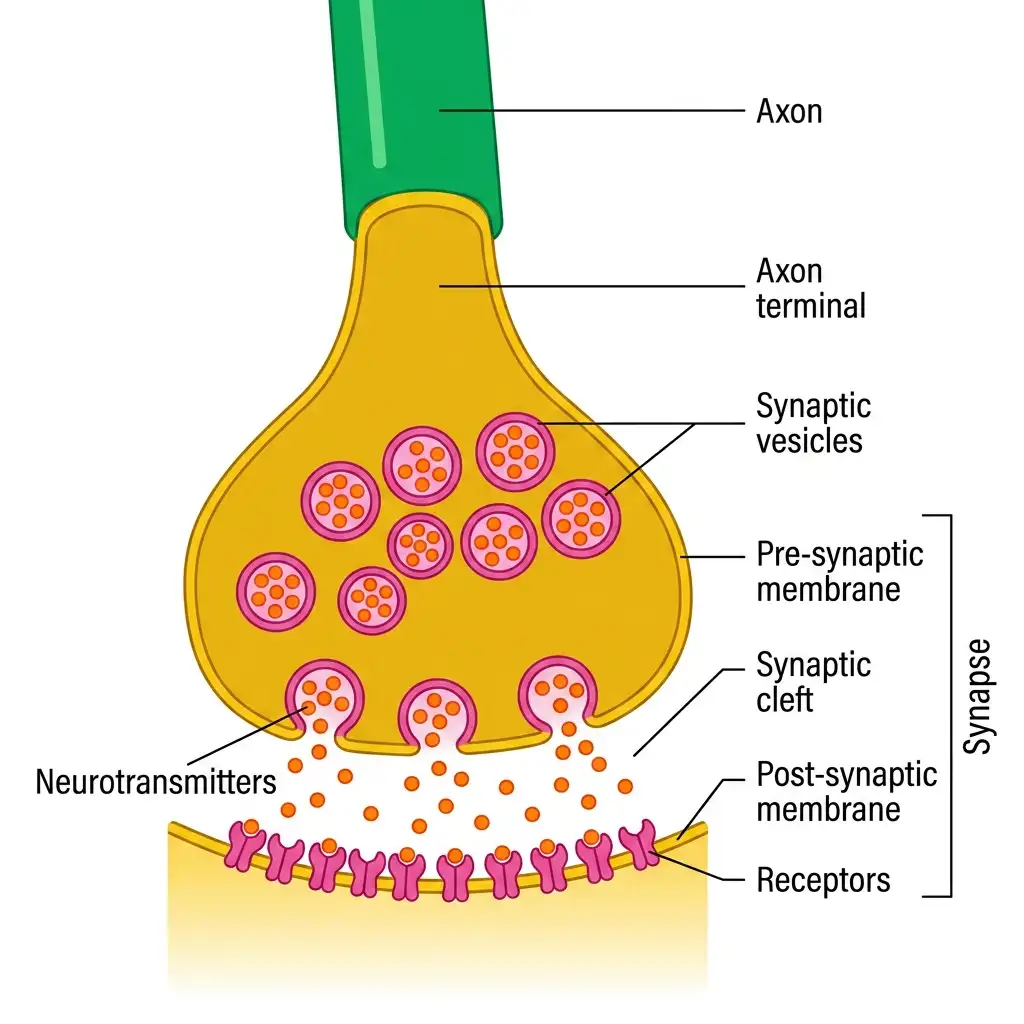
Synapse Structure: Shows how electrical signals jump across a synaptic cleft using neurotransmitter-filled vesicles.
🔬 Detailed Anatomical Description
A nerve impulse is transmitted between neurons across a junction called a synapse. At a chemical synapse, an action potential reaching the axon terminal triggers calcium influx, causing synaptic vesicles to fuse with the pre-synaptic membrane. Vesicles release neurotransmitters into the synaptic cleft by exocytosis. These molecules diffuse across the cleft and bind to specific receptors on the post-synaptic membrane, generating a new electrical potential.
XIII. CENTRAL NEURAL SYSTEM (CNS)
| FEATURE | DETAIL |
|---|---|
| CNS consists of | Brain + spinal cord ⭐⭐⭐ |
| Brain role | Central information processing organ; command and control system ⭐⭐⭐ |
Major Functions of Brain ⭐⭐⭐
- Controls voluntary movements
- Maintains balance
- Controls vital involuntary organ functions (lungs, heart, kidneys)
- Thermoregulation
- Hunger and thirst
- Circadian rhythms
- Activities of several endocrine glands
- Behaviour
- Processing of vision, hearing, speech, memory, intelligence, emotions, thoughts
XIV. PROTECTION OF BRAIN / SKELETAL ARTICULATIONS
| FEATURE | DETAIL |
|---|---|
| Brain protected externally by | Skull ⭐⭐⭐ |
| Covering inside skull | Cranial meninges ⭐⭐⭐ |
Cranial Meninges ⭐⭐⭐
| LAYER | POSITION | DETAIL |
|---|---|---|
| Dura mater | Outer | Tough outer layer ⭐⭐⭐ |
| Arachnoid | Middle | Very thin middle layer ⭐⭐⭐ |
| Pia mater | Inner | In contact with brain tissue ⭐⭐⭐ |
NEET 2026: The occipital bone of the skull is articulated with the first vertebra.
NEET 2026: In humans, the number of cervical vertebrae is seven.
NEET 2026: All ribs except the last 2 pairs are bicephalic.
NEET 2026: The joint between any two adjoining vertebrae is a cartilaginous joint.
XV. MAJOR PARTS OF BRAIN
| MAJOR DIVISION | COMPONENTS |
|---|---|
| Forebrain | Cerebrum, Thalamus, Hypothalamus ⭐⭐⭐ |
| Midbrain | Corpora quadrigemina region etc. ⭐⭐⭐ |
| Hindbrain | Pons, Cerebellum, Medulla oblongata ⭐⭐⭐ |
Sagittal Section of Human Brain
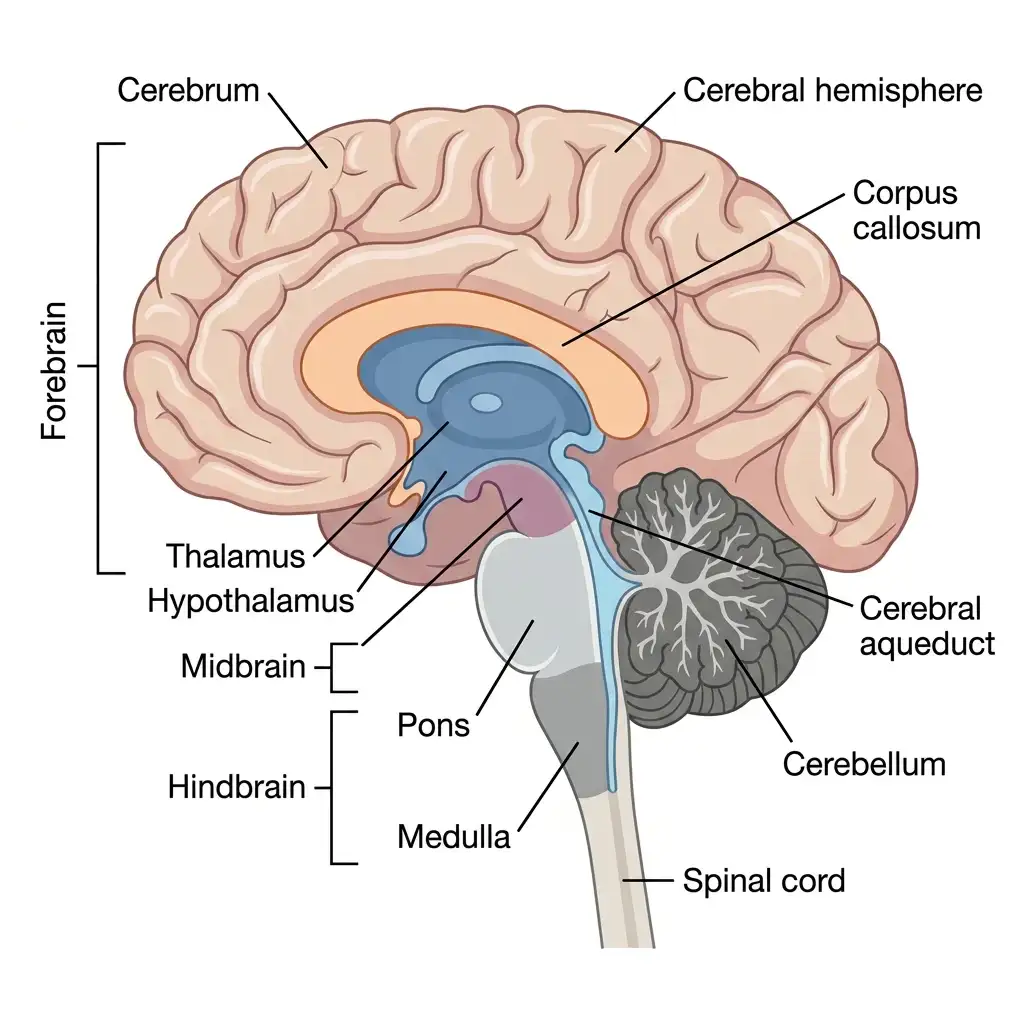
Sectional View of Human Brain: Depicts sagittal coordinates of brain anatomy, detailing cerebrum, corpus callosum, pons, and cerebellum.
🔬 Detailed Anatomical Description
The human brain is divided into: (1) Forebrain: Consists of the cerebrum (divided into hemispheres by the corpus callosum), thalamus (sensory relay center), and hypothalamus (homeostasis center); (2) Midbrain: Located between forebrain and hindbrain, containing the cerebral aqueduct; (3) Hindbrain: Comprises the pons (pneumotaxic center), cerebellum (coordination and balance), and medulla oblongata (cardiac and respiratory centers) leading to the spinal cord.
XVI. FOREBRAIN
A CEREBRUM
| FEATURE | DETAIL |
|---|---|
| Cerebrum | Major part of human brain ⭐⭐⭐ |
| Also | Most developed part of human brain ⭐⭐⭐ |
| Deep cleft divides it into | Left and right cerebral hemispheres ⭐⭐⭐ |
| Hemispheres connected by | Corpus callosum ⭐⭐⭐ |
NEET 2015, 2018, 2024: Corpus callosum connects the two cerebral hemispheres.
Corpus Callosum ⭐⭐⭐
| FEATURE | DETAIL |
|---|---|
| Nature | Thick band / tract of nerve fibres ⭐⭐⭐ |
| Function | Allows communication between two cerebral hemispheres ⭐⭐⭐ |
B CEREBRAL CORTEX
| FEATURE | DETAIL |
|---|---|
| Outer layer covering hemispheres | Cerebral cortex ⭐⭐⭐ |
| Appearance | Thrown into prominent folds ⭐⭐ |
| Grey matter | Cerebral cortex; contains many neuron cell bodies ⭐⭐⭐ |
| White matter | Inner part of cerebrum; due to myelinated fibres ⭐⭐⭐ |
Functional Areas of Cerebral Cortex ⭐⭐⭐
| AREA | FUNCTION |
|---|---|
| Motor areas | Motor functions ⭐⭐⭐ |
| Sensory areas | Sensory functions ⭐⭐⭐ |
| Association areas | Neither purely motor nor sensory ⭐⭐⭐ |
Association Areas ⭐⭐⭐
| FUNCTION | DETAIL |
|---|---|
| Memory | ⭐⭐⭐ |
| Communication | ⭐⭐⭐ |
| Intersensory association | ⭐⭐⭐ |
| Higher functions | Calculation, contemplation, language comprehension ⭐⭐ |
C THALAMUS
| FEATURE | DETAIL |
|---|---|
| Position | Cerebrum wraps around thalamus ⭐⭐ |
| Function | Major coordinating centre for sensory and motor signalling ⭐⭐⭐ |
D HYPOTHALAMUS
| FEATURE | DETAIL |
|---|---|
| Position | Lies at the base of thalamus ⭐⭐⭐ |
| Controls | Body temperature, urge of eating, urge of drinking ⭐⭐⭐ |
| Contains | Groups of neurosecretory cells ⭐⭐⭐ |
| These cells secrete | Hypothalamic hormones / releasing hormones ⭐⭐⭐ |
NEET 2014, 2018, 2019, 2023, 2024: Hypothalamus controls thermoregulation, hunger, thirst.
Injury to hypothalamus directly affects body temperature regulation.
E LIMBIC LOBE / LIMBIC SYSTEM
| FEATURE | DETAIL |
|---|---|
| Formed by | Inner parts of cerebral hemispheres + deep structures like amygdala, hippocampus etc. ⭐⭐⭐ |
| Works along with | Hypothalamus ⭐⭐⭐ |
| Functions | Regulation of sexual behaviour, emotional reactions, motivation ⭐⭐⭐ |
Emotional Reactions Regulated ⭐⭐⭐
- Excitement
- Pleasure
- Rage
- Fear
Additional NCERT Summary Point ⭐⭐
- Olfaction
- Autonomic responses
NEET 2023: Limbic system + hypothalamus regulate sexual behaviour and emotions.
TRAP: Limbic system does not control physical movement.
XVII. MIDBRAIN
| FEATURE | DETAIL |
|---|---|
| Location | Between thalamus / hypothalamus of forebrain and pons of hindbrain ⭐⭐⭐ |
| Canal passing through it | Cerebral aqueduct ⭐⭐ |
| Dorsal part contains | Four rounded swellings called corpora quadrigemina ⭐⭐⭐ |
Additional Function ⭐⭐
| FEATURE | DETAIL |
|---|---|
| Midbrain function | Receives and integrates visual, tactile and auditory inputs ⭐⭐ |
XVIII. HINDBRAIN
| COMPONENT | DETAIL |
|---|---|
| Pons | Fibre tracts connecting different brain regions ⭐⭐⭐ |
| Cerebellum | Highly convoluted surface for accommodating more neurons ⭐⭐⭐ |
| Medulla oblongata | Connected to spinal cord; contains centres for vital reflexes ⭐⭐⭐ |
XIX. PONS
| FEATURE | DETAIL |
|---|---|
| Main role | Contains fibre tracts interconnecting different regions of brain ⭐⭐⭐ |
| Additional NEET fact | Contains pneumotaxic centre ⭐⭐ |
| Pneumotaxic centre function | Alters respiratory rate by reducing duration of inspiration ⭐⭐ |
NEET 2022, 2024: Pons has important respiratory role via pneumotaxic centre.
XX. CEREBELLUM
| FEATURE | DETAIL |
|---|---|
| Surface | Very highly convoluted ⭐⭐⭐ |
| Reason | Provides additional space for many more neurons ⭐⭐⭐ |
| Main functions | Regulation of posture, balance, and coordination ⭐⭐⭐ |
TRAP: Cerebellum does not control language comprehension.
XXI. MEDULLA OBLONGATA
| FEATURE | DETAIL |
|---|---|
| Position | Connected to spinal cord ⭐⭐⭐ |
| Contains centres controlling | Respiration, cardiovascular reflexes, gastric secretions ⭐⭐⭐ |
NEET 2015, 2018, 2024: Medulla controls respiration, cardiovascular reflexes, gastric secretion.
XXII. BRAIN STEM
| FEATURE | DETAIL |
|---|---|
| Brain stem consists of | Midbrain + Pons + Medulla oblongata ⭐⭐⭐ |
| Function | Forms connection between brain and spinal cord ⭐⭐⭐ |
NEET 2023, 2024: Brain stem = midbrain + pons + medulla.
TRAP: Cerebrum is NOT part of brain stem.
XXIII. RAPID REVISION — KEY COMPARISON TABLES
TABLE 1: CNS vs PNS ⭐⭐⭐
| FEATURE | CNS | PNS |
|---|---|---|
| Full form | Central Neural System | Peripheral Neural System |
| Components | Brain + spinal cord | All nerves associated with CNS |
| Role | Information processing and control | Carries impulses to and from CNS |
TABLE 2: Afferent vs Efferent Fibres ⭐⭐⭐
| FEATURE | AFFERENT | EFFERENT |
|---|---|---|
| Direction | Tissue / organ → CNS | CNS → peripheral organ / tissue |
| Role | Sensory input | Motor / regulatory output |
TABLE 3: Somatic vs Autonomic Nervous System ⭐⭐⭐
| FEATURE | SOMATIC | AUTONOMIC |
|---|---|---|
| Target | Skeletal muscles | Involuntary organs + smooth muscles |
| Type of control | Voluntary | Involuntary |
TABLE 4: Parts of a Neuron ⭐⭐⭐
| PART | FUNCTION / FEATURE |
|---|---|
| Cell body | Organelles + Nissl bodies |
| Dendrites | Carry impulses toward cell body |
| Axon | Carries impulses away from cell body |
| Synaptic knob | Contains neurotransmitter vesicles |
TABLE 5: Types of Neurons ⭐⭐⭐
| TYPE | STRUCTURE | EXAMPLE |
|---|---|---|
| Multipolar | 1 axon + many dendrites | Cerebral cortex |
| Bipolar | 1 axon + 1 dendrite | Retina |
| Unipolar | Only 1 axon | Embryonic stage |
TABLE 6: Myelinated vs Non-myelinated Fibres ⭐⭐⭐
| FEATURE | MYELINATED | NON-MYELINATED |
|---|---|---|
| Myelin sheath | Present | Absent |
| Formed by Schwann cells | Yes | Schwann cell present but sheath absent |
| Node of Ranvier | Present | Absent |
| Common location | Spinal and cranial nerves | Autonomic and some somatic fibres |
TABLE 7: Resting Potential vs Action Potential ⭐⭐⭐
| FEATURE | RESTING POTENTIAL | ACTION POTENTIAL |
|---|---|---|
| Membrane state | Polarised | Depolarised |
| Outer surface | Positive | Negative |
| Inner surface | Negative | Positive |
| Major ion movement | Na⁺/K⁺ pump maintained | Rapid Na⁺ influx |
TABLE 8: Electrical vs Chemical Synapse ⭐⭐⭐
| FEATURE | ELECTRICAL SYNAPSE | CHEMICAL SYNAPSE |
|---|---|---|
| Synaptic cleft | Absent / negligible | Present |
| Transmission | Direct electrical current | Neurotransmitter mediated |
| Speed | Faster | Slower |
| Occurrence | Rare | Common |
TABLE 9: Brain Divisions ⭐⭐⭐
| DIVISION | COMPONENTS |
|---|---|
| Forebrain | Cerebrum, Thalamus, Hypothalamus |
| Midbrain | Corpora quadrigemina region |
| Hindbrain | Pons, Cerebellum, Medulla |
TABLE 10: Brain Regions and Functions ⭐⭐⭐
| PART | FUNCTION |
|---|---|
| Cerebrum | Higher functions, memory, thought, language, voluntary acts |
| Thalamus | Coordination of sensory and motor signalling |
| Hypothalamus | Thermoregulation, hunger, thirst, hormones |
| Limbic system | Emotions, sexual behaviour, motivation |
| Midbrain | Integrates visual / tactile / auditory inputs |
| Pons | Fibre tracts; pneumotaxic centre |
| Cerebellum | Balance, posture, coordination |
| Medulla | Respiration, cardiovascular reflexes, gastric secretion |
TABLE 11: Cranial Meninges ⭐⭐⭐
| LAYER | POSITION |
|---|---|
| Dura mater | Outer |
| Arachnoid | Middle |
| Pia mater | Inner |
TABLE 12: Important Transport Facts ⭐⭐⭐
| FEATURE | DETAIL |
|---|---|
| Na⁺/K⁺ pump | 3 Na⁺ out, 2 K⁺ in |
| Receptor for neurotransmitter | Post-synaptic membrane |
| Synaptic vesicles | Present in synaptic knob |
| Causing depolarisation | Rapid Na⁺ influx |
XXIV. KEY NUMBERS / FACTS — QUICK REFERENCE
| PARAMETER / FACT | VALUE / DETAIL |
|---|---|
| Main parts of neuron | 3 — cell body, dendrites, axon |
| Divisions of human neural system | 2 — CNS and PNS |
| Main divisions of brain | 3 — forebrain, midbrain, hindbrain |
| Na⁺/K⁺ pump ratio | 3 Na⁺ out : 2 K⁺ in |
| Brain stem components | Midbrain + Pons + Medulla |
| Cranial meninges layers | 3 — dura, arachnoid, pia |
| Types of neuron | 3 — multipolar, bipolar, unipolar |
| Types of synapse | 2 — electrical, chemical |
XXV. COMMON EXAM TRAPS — QUICK REFERENCE
| TRAP / QUESTION | CORRECT ANSWER |
|---|---|
| Neural system gives slow coordination? | No — quick point-to-point coordination |
| Endocrine system provides point-to-point connection? | No — chemical integration via hormones |
| CNS consists of only brain? | No — brain + spinal cord |
| Afferent fibres carry impulses from CNS to organs? | No — from organs / tissues to CNS |
| Efferent fibres carry impulses to CNS? | No — from CNS to tissues / organs |
| Somatic NS supplies smooth muscles? | No — skeletal muscles |
| Autonomic NS supplies skeletal muscle? | No — involuntary organs, smooth muscles |
| Dendrites carry impulses away from cell body? | No — towards cell body |
| Axon carries impulse toward cell body? | No — away from cell body |
| Nissl bodies present in axon? | No — in cell body and dendrites |
| Multipolar neuron found in retina? | No — cerebral cortex |
| Bipolar neuron found in cortex? | No — retina |
| Myelin in PNS formed by oligodendrocytes? | No — Schwann cells |
| Myelin in CNS formed by Schwann cells? | No — oligodendrocytes |
| Resting membrane more permeable to Na⁺? | No — more permeable to K⁺ |
| Na⁺/K⁺ pump moves 2 Na⁺ out and 3 K⁺ in? | No — 3 Na⁺ out, 2 K⁺ in |
| Resting outer membrane negative? | No — outer positive, inner negative |
| Action potential at rest? | No — resting potential at rest |
| Depolarisation caused by K⁺ influx? | No — rapid Na⁺ influx |
| Electrical synapse slower than chemical? | No — electrical is faster |
| Neurotransmitter receptors present on presynaptic membrane? | No — on post-synaptic membrane |
| Synaptic knob contains hormones? | No — neurotransmitter vesicles |
| Cerebrum is part of brain stem? | No |
| Brain stem = ? | Midbrain + Pons + Medulla |
| Corpus callosum connects cerebrum and cerebellum? | No — left and right cerebral hemispheres |
| Cerebrum most developed part of brain? | Yes |
| Cerebral cortex = white matter? | No — grey matter |
| White matter outside and cortex inside? | No — cortex outside, white matter inside |
| Association areas are sensory / motor only? | No — complex functions like memory, communication |
| Hypothalamus controls temperature, hunger, thirst? | Yes |
| Hypothalamus secretes releasing hormones? | Yes, via neurosecretory cells |
| Limbic system controls physical movement? | No |
| Limbic system + hypothalamus regulate emotions and sex behaviour? | Yes |
| Cerebellum controls language comprehension? | No |
| Cerebellum role? | Posture, balance, coordination |
| Medulla controls respiration and cardiovascular reflexes? | Yes |
| Pons contains fibre tracts? | Yes |
| Inner meningeal layer in contact with brain tissue? | Pia mater |