I. ANIMAL TISSUES — OVERVIEW & CLASSIFICATION
In multicellular animals, a group of similar cells along with intercellular substances perform a specific function. Such an organization is called a tissue. All complex animals consist of only four basic types of tissues which are organized in specific proportions and patterns to form organs such as stomach, lung, heart, and kidney.
| TISSUE TYPE | EMBRYONIC ORIGIN | PRIMARY FUNCTION | EXAMPLES & LOCATIONS |
|---|---|---|---|
| Epithelial Tissue | Ectoderm, Mesoderm, or Endoderm | Protection, secretion, absorption, excretion, sensory reception | Skin epidermis, lining of digestive & respiratory tracts, glands |
| Connective Tissue | Mesoderm | Binding, structural support, protection, insulation, transport | Areolar tissue, adipose tissue, tendons, ligaments, cartilage, bone, blood |
| Muscular Tissue | Mesoderm | Locomotion and movement of body parts via contraction | Skeletal muscle, smooth (visceral) muscle, cardiac muscle |
| Neural Tissue | Ectoderm | Control, coordination, generation & conduction of nerve impulses | Brain, spinal cord, peripheral nerves, ganglia |
NEET 2018, 2023 (reinforced): Epithelial tissue is the only animal tissue that can originate from all three embryonic germ layers (Ectoderm, Mesoderm, and Endoderm), whereas Connective and Muscular tissues originate exclusively from Mesoderm, and Neural tissue from Ectoderm.
II. EPITHELIAL TISSUE — SIMPLE, COMPOUND & SPECIALISED EPITHELIA
Epithelial Tissue (Epithelium): Has a free surface which faces either a body fluid or the outside environment, thus providing a covering or a lining for some part of the body. The cells are compactly packed with little intercellular matrix. Epithelium rests on a non-cellular basement membrane.
A SIMPLE EPITHELIUM (SINGLE LAYER OF CELLS)
| TYPE | STRUCTURAL FEATURES | NCERT LOCATIONS / EXAMPLES |
|---|---|---|
| Simple Squamous Epithelium | Single thin layer of flattened cells with irregular boundaries (Pavement epithelium) | Walls of blood vessels (endothelium), air sacs of lungs (alveoli), Bowman's capsule of nephron ⭐⭐⭐ |
| Simple Cuboidal Epithelium | Single layer of cube-like cells; nucleus is spherical and centrally located | Ducts of glands, tubular parts of nephrons in kidneys (PCT has brush border microvilli), germinal epithelium of ovary/testis ⭐⭐⭐ |
| Simple Columnar Epithelium | Single layer of tall and slender cells; basal oval nuclei; free surface may have microvilli | Lining of stomach and intestine (secretion and absorption) ⭐⭐⭐ |
| Ciliated Epithelium | Cuboidal or columnar cells bearing cilia on their free surface; move particles/mucus in specific direction | Inner surface of hollow organs like bronchioles and Fallopian tubes (oviducts) ⭐⭐⭐ |
| Pseudostratified Epithelium | Appears multi-layered due to nuclei at different levels, but all cells rest on single basement membrane | Lining of trachea, larger bronchi, nasal cavities ⭐⭐⭐ |
B COMPOUND & GLANDULAR EPITHELIUM
| CATEGORY | DESCRIPTION | EXAMPLES / LOCATIONS |
|---|---|---|
| Compound Epithelium | Multi-layered (stratified); limited role in secretion/absorption; main function is protection against chemical and mechanical stresses | Dry surface of skin (keratinized), moist surface of buccal cavity, pharynx, inner lining of ducts of salivary glands and pancreatic ducts ⭐⭐⭐ |
| Transitional Epithelium | Stretchable stratified epithelium without basement membrane in stretched state | Lining of urinary bladder and ureters (urothelium) ⭐⭐⭐ |
| Unicellular Glands | Isolated glandular epithelial cells | Goblet cells of alimentary canal (secrete mucus) ⭐⭐⭐ |
| Multicellular Glands | Cluster of glandular epithelial cells | Salivary glands, gastric glands, tear glands ⭐⭐⭐ |
C SPECIALISED CELL JUNCTIONS
| JUNCTION TYPE | EXACT NCERT FUNCTION |
|---|---|
| Tight Junctions | Help to stop substances from leaking across a tissue ⭐⭐⭐ |
| Adhering Junctions (Desmosomes) | Perform cementing to keep neighbouring cells together ⭐⭐⭐ |
| Gap Junctions | Facilitate cells to communicate with each other by connecting the cytoplasm of adjoining cells, for rapid transfer of ions, small molecules, and sometimes big molecules ⭐⭐⭐ |
NEET 2019, 2021, 2023 (reinforced): The proximal convoluted tubule (PCT) of nephron in kidney has simple cuboidal epithelium with a prominent brush border of microvilli which increases surface area for reabsorption. Fallopian tubes and bronchioles are lined by ciliated epithelium.
III. CONNECTIVE TISSUE — LOOSE, DENSE & SPECIALISED
Connective Tissue: Most abundant and widely distributed in the body of complex animals. Named connective tissues because of their special function of linking and supporting other tissues/organs of the body. In all connective tissues except blood, cells secrete fibres of structural proteins called collagen or elastin which provide strength, elasticity, and flexibility.
| CLASS | TISSUE TYPE | KEY FEATURES & NCERT LOCATIONS |
|---|---|---|
| Loose Connective Tissue (Cells & fibres loosely arranged in semi-fluid ground substance) |
Areolar Tissue | Present beneath skin; serves as support framework for epithelium. Contains fibroblasts (secrete fibres), macrophages (phagocytic), and mast cells (secrete histamine, serotonin, heparin) ⭐⭐⭐ |
| Adipose Tissue | Located mainly beneath skin; specialized to store fat. Excess nutrients are converted into fats and stored in adipocytes ⭐⭐⭐ | |
| Dense Connective Tissue (Fibres and fibroblasts compactly packed) |
Dense Regular Connective Tissue | Collagen fibres present in rows between many parallel bundles of fibres. • Tendons: Connect skeletal muscle to bone. • Ligaments: Connect bone to bone ⭐⭐⭐ |
| Dense Irregular Connective Tissue | Fibroblasts and many fibres (mostly collagen) oriented differently. Present in the dermis of skin ⭐⭐⭐ | |
| Specialised Connective Tissue | Cartilage | Intercellular material is solid, pliable, and resists compression. Cells (chondrocytes) enclosed in small cavities (lacunae). Found in tip of nose, outer ear joints (pinna), between vertebral bones, limbs/hands in adults ⭐⭐⭐ |
| Bone | Hard, non-pliable ground substance rich in calcium salts and collagen fibres giving strength. Osteocytes present in spaces called lacunae. Bone marrow in some bones is the site of hematopoiesis ⭐⭐⭐ | |
| Blood | Fluid connective tissue consisting of plasma, red blood cells (RBCs), white blood cells (WBCs), and platelets. Only connective tissue where fibres are NOT secreted ⭐⭐⭐ |
NEET 2016, 2020, 2023 (reinforced): Tendon connects muscle to bone, while Ligament connects bone to bone. Both are examples of Dense Regular Connective Tissue. Dermis of skin is an example of Dense Irregular Connective Tissue.
IV. MUSCULAR TISSUE & NEURAL TISSUE
Muscular Tissue: Consists of long, cylindrical fibres arranged in parallel arrays composed of fine fibrils called myofibrils. Contract (shorten) in response to stimulation and relax (lengthen) in a coordinated fashion to produce movement.
| FEATURE | SKELETAL / STRIATED MUSCLE | SMOOTH / VISCERAL MUSCLE | CARDIAC MUSCLE |
|---|---|---|---|
| Location | Attached to skeletal bones (e.g., biceps, triceps) | Walls of internal hollow organs (blood vessels, stomach, intestine) | Wall of the heart exclusively ⭐⭐⭐ |
| Striations | Present (prominent light & dark bands) | Absent (smooth / non-striated appearance) | Present (faint cross-striations) |
| Cell Shape | Long, cylindrical, unbranched | Spindle-shaped (fusiform), tapering ends | Short, cylindrical, branched network |
| Nucleus | Multinucleated (syncytial), peripheral | Uninucleated, centrally located | Uninucleated, centrally located |
| Control | Voluntary (under conscious control) | Involuntary (not under conscious control) | Involuntary (myogenic rhythmic contraction) |
| Intercalated Discs | Absent | Absent | Present (communication junctions / adhesion discs allow synchronized wave of contraction) ⭐⭐⭐ |
NEURAL TISSUE
Neural Tissue: Exerts the greatest control over the body's responsiveness to changing conditions. Consists of two distinct cell types:
- Neurons: Excitable cells that are the structural and functional units of neural system. Capable of generating and conducting electrical action potentials along their axon.
- Neuroglial Cells: Non-excitable supporting cells that protect and support neurons. Neuroglia make up more than one-half the volume of neural tissue in our body.
NEET 2017, 2022 (reinforced): Intercalated discs are unique to cardiac muscle and act as communication junctions so that when one cell receives a signal to contract, its neighbours are also stimulated to contract. Neuroglial cells constitute >50% of neural tissue volume.
V. ORGAN & ORGAN SYSTEM — MULTICELLULAR ARCHITECTURE
In multicellular organisms, basic tissues group together to form organs, and organs associate to form organ systems. This organization is essential for efficient division of labour and survival of the organism.
In the study of structural organisation in animals, three representative model organisms are classically examined across invertebrate and vertebrate phyla:
- Earthworm (Pheretima): Representative invertebrate of Phylum Annelida.
- Cockroach (Periplaneta americana): Representative invertebrate of Phylum Arthropoda.
- Frog (Rana tigrina): Representative vertebrate of Phylum Chordata, Class Amphibia.
VI. FROG — RANA TIGRINA • A. GENERAL CHARACTERISTICS & CLASSIFICATION
Frog (Rana tigrina): Frogs are amphibious vertebrates that can live both on land and in freshwater. The most common species of frog found in India is Rana tigrina (Indian bullfrog).
| TAXONOMIC CATEGORY | CLASSIFICATION OF FROG |
|---|---|
| Kingdom | Animalia |
| Phylum | Chordata (Notochord present in embryonic stage; dorsal hollow nerve cord) |
| Subphylum | Vertebrata (Craniata — skull present; vertebral column replaces notochord) |
| Class | Amphibia (Dual life — water and terrestrial habitat; pentadactyl limbs) |
| Genus | Rana |
| Species | tigrina (Binomial citation: Rana tigrina Daud.) |
GENERAL BIOLOGICAL FEATURES & ADAPTATIONS
| FEATURE | SCIENTIFIC EXPLANATION (NCERT VERBATIM POINTS) |
|---|---|
| Poikilothermic (Cold-blooded) | Frogs do not have a constant body temperature; their body temperature fluctuates with the temperature of the environment. They are poikilotherms or ectotherms ⭐⭐⭐ |
| Camouflage & Mimicry | Ability to change skin color to blend with surroundings and hide from predators (camouflage). The protective color change is also termed metachrosis ⭐⭐⭐ |
| Aestivation (Summer Sleep) | During peak summer season, frogs take shelter in deep underground burrows to escape extreme heat and desiccation ⭐⭐⭐ |
| Hibernation (Winter Sleep) | During severe winter season, frogs burrow deep into soil or mud to escape extreme cold temperatures and enter metabolic dormancy ⭐⭐⭐ |
| Nucleated RBCs | Red blood cells (erythrocytes) of frogs are oval, biconvex, and nucleated, containing red pigment haemoglobin (in contrast to human RBCs which are enucleated) ⭐⭐⭐ |
NEET 2026 context / reinforced: Frog RBCs are nucleated. Unlike mammalian erythrocytes which lose their nucleus upon maturity, frog erythrocytes retain a functional oval nucleus throughout their lifespan in circulation.
VII. FROG — RANA TIGRINA • B. MORPHOLOGY (EXTERNAL FEATURES & SEXUAL DIMORPHISM)
External Morphology of Frog: The skin is smooth and slippery due to the presence of mucus. The skin is always maintained in a moist condition. The dorsal side of body is generally olive green with dark irregular spots, while the ventral side is uniformly pale yellow. The frog never drinks water but absorbs it through the skin.
The body of a frog is divisible into two regions only: Head and Trunk. Neck and tail are completely absent in the adult frog.
| BODY PART | DETAILED NCERT MORPHOLOGY |
|---|---|
| Head Region | • Anteriorly snout is roughly pointed. • External nostrils present above mouth. • Eyes: Bulging, covered by a translucent nictitating membrane that protects the eyeball while in water. • Tympanum: A membranous eardrum present on either side of eyes, receiving sound signals (external ear is absent) ⭐⭐⭐ |
| Forelimbs | Short, end in four digits. Absorb shock when landing after a leap. No webbing present between digits ⭐⭐⭐ |
| Hindlimbs | Much larger and more muscular than forelimbs; end in five digits with webbed skin between toes which acts as a paddle during swimming and assists in jumping ⭐⭐⭐ |
| Sexual Dimorphism | Male frogs can be distinguished from female frogs by the presence of: 1. Vocal sacs: Sound-producing resonators under neck. 2. Copulatory pad: Present on the first digit of the forelimb (index finger), used to clasp female during amplexus. Both vocal sacs and copulatory pads are ABSENT in female frogs. ⭐⭐⭐ |
NEET 2011, 2016, 2026 (reinforced): Male frogs can be distinguished from female frogs due to the presence of vocal sacs and copulatory pads (on the first digit of the forelimb). Remember: forelimbs have 4 digits, hindlimbs have 5 digits!
Frog External Anatomy
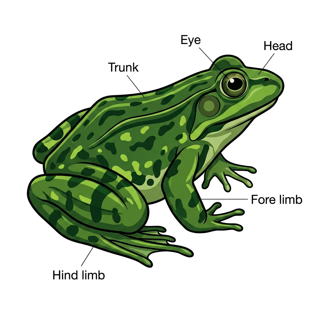
External Anatomy of Frog: Highlights the specialized body divisions and jointed limbs of Rana tigrina.
🔬 Detailed Anatomical Description
Rana tigrina is the most common frog species. Its body is divided into: (1) Head: Features eyes covered by a nictitating membrane, and a tympanum ear drum; (2) Trunk: Accommodates internal visceral organs. The limbs are highly specialized: the short forelimbs contain four digits, and the long, muscular hindlimbs end in five webbed digits designed for jumping and swimming.
VIII. FROG — RANA TIGRINA • C. ANATOMY (1) — DIGESTIVE SYSTEM
Digestive System of Frog: Consists of the alimentary canal and digestive glands. The alimentary canal is short because frogs are carnivores, and hence the length of the intestine is reduced.
| ORGAN / GLAND | FUNCTION & NCERT DIGESTIVE MECHANISM |
|---|---|
| Mouth & Tongue | Mouth opens into buccal cavity. Prey is captured by a bilobed, sticky protrusible tongue ⭐⭐⭐ |
| Oesophagus & Stomach | Oesophagus is a short tube opening into stomach. Stomach walls secrete HCl and gastric juice. Partially digested acidic food in stomach is called chyme ⭐⭐⭐ |
| Liver & Gall Bladder | Liver is a trilobed dark brown gland that secretes bile which is stored in the light-green gall bladder. Bile emulsifies fat ⭐⭐⭐ |
| Pancreas | Digestive gland that secretes pancreatic juice containing digestive enzymes (trypsin, amylase, lipase) ⭐⭐⭐ |
| Intestine (Duodenum & Ileum) | Duodenum receives bile and pancreatic juice through a common hepato-pancreatic duct. Bile emulsifies fats; pancreatic enzymes digest carbohydrates and proteins. Final digestion and absorption occur in intestine via finger-like folds called villi and microvilli ⭐⭐⭐ |
| Rectum & Cloaca | Undigested waste moves into wide rectum and opens into cloaca, exiting exteriorly via cloacal aperture ⭐⭐⭐ |
Frog Digestive System
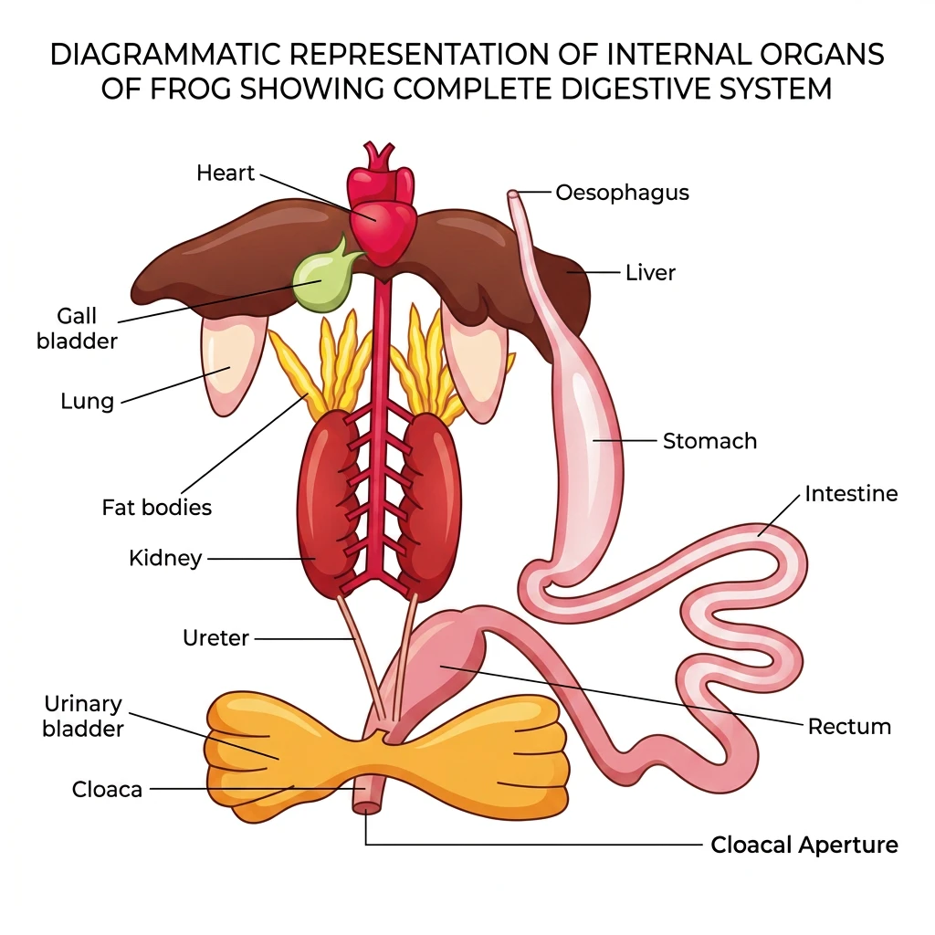
Frog Visceral Organs: Displays the complete digestive tract, heart, and accessory organs (liver and gall bladder).
🔬 Detailed Anatomical Description
The internal digestive system of a frog consists of the alimentary canal and accessory glands: (1) Alimentary canal: Begins at mouth, leading down to the short esophagus, muscular stomach, coiled intestine, wide rectum, and terminates at the cloaca and cloacal aperture; (2) Glands: The trilobed dark-brown liver wraps the red heart and secretes bile stored in the light-green gall bladder, while the pancreas secretes pancreatic juice.
IX. FROG — RANA TIGRINA • C. ANATOMY (2) — RESPIRATORY SYSTEM
Respiratory System of Frog: Frogs respire on land and in water by two different methods. They exhibit cutaneous respiration, buccal respiration, and pulmonary respiration depending on their habitat and activity state.
| RESPIRATORY MODE | MECHANISM & HABITAT SPECIFICITY |
|---|---|
| Cutaneous Respiration (Skin) | In water, skin acts as the aquatic respiratory organ. Dissolved oxygen in water is exchanged through the moist skin by simple diffusion. Skin also acts as the sole respiratory organ during aestivation and hibernation ⭐⭐⭐ |
| Buccal Respiration (Buccal Cavity) | On land, the buccal cavity lining is richly vascularized; air enters through external nostrils into the buccal cavity where exchange of O₂ and CO₂ takes place across the mucous lining ⭐⭐⭐ |
| Pulmonary Respiration (Lungs) | On land, respiration also occurs by lungs. Lungs are a pair of elongated, pink, sac-like structures present in the upper part of the trunk region (thorax). Air enters through nostrils into the buccal cavity and then into lungs ⭐⭐⭐ |
NEET 2026 / RE-NEET 2026 context: In frogs, respiration in water is by skin (cutaneous); on land by skin, buccal cavity and lungs (statement true for both environments).
RE-NEET 2026: In water, frogs respire using skin. Remember: skin respiration operates in water AND on land AND during seasonal dormancy!
X. FROG — RANA TIGRINA • C. ANATOMY (3) — CIRCULATORY SYSTEM
Circulatory System of Frog: Frog has a well-developed closed type of circulatory system with incomplete double circulation. It also possesses a lymphatic system consisting of lymph, lymph channels, and lymph nodes.
A HEART & SPECIAL ACCESSIORY STRUCTURES
| STRUCTURE | ANATOMICAL DETAIL |
|---|---|
| Heart Chambers | Three-chambered muscular heart situated in the upper part of body cavity: two atria (left and right) and one ventricle. Covered by a double-walled pericardium ⭐⭐⭐ |
| Sinus Venosus | A triangular chamber attached dorsally to the right atrium; receives deoxygenated blood through major veins called caval veins (vena cava) ⭐⭐⭐ |
| Conus Arteriosus | A sac-like muscular structure on the ventral side of the heart into which the single ventricle opens ⭐⭐⭐ |
B SPECIAL PORTAL SYSTEMS & BLOOD
| SYSTEM / COMPONENT | SCIENTIFIC DEFINITION |
|---|---|
| Hepatic Portal System | Special venous connection between liver and intestine ⭐⭐⭐ |
| Renal Portal System | Special venous connection between kidney and lower part of body (hindlimbs) ⭐⭐⭐ |
| Blood Elements & RBCs | Blood consists of plasma and formed elements: RBCs (erythrocytes), WBCs (leucocytes), and platelets. • Frog RBCs are nucleated, oval, and biconvex containing red pigment haemoglobin ⭐⭐⭐ |
NEET 2026 context: Hepatic portal system = special venous connection between liver and intestine.
NEET 2026 context: Renal portal system = special venous connection between kidney and lower part of body.
NEET 2013, 2026 context (reinforced): Frog RBCs are nucleated!
XI. FROG — RANA TIGRINA • C. ANATOMY (4) — EXCRETORY SYSTEM
Excretory System of Frog: Eliminates nitrogenous wastes from the body. Consists of a pair of kidneys, ureters, cloaca, and urinary bladder.
| STRUCTURE | NCERT ANATOMICAL DETAIL |
|---|---|
| Kidneys & Nephrons | A pair of compact, dark red, bean-like kidneys situated posteriorly on both sides of vertebral column. Each kidney contains numerous structural and functional units called uriniferous tubules or nephrons ⭐⭐⭐ |
| Ureters in Male Frog | Two ureters emerge from kidneys. In males, they act as urinogenital ducts which carry both urine and spermatozoa, opening into the cloaca ⭐⭐⭐ |
| Ureters in Female Frog | In females, the ureters and oviducts open independently into the cloaca (ureters carry urine only) ⭐⭐⭐ |
| Urinary Bladder & Cloaca | Thin-walled urinary bladder is situated ventral to rectum and opens into cloaca. Cloaca is a small median chamber that receives urine, fecal matter, and sex cells ⭐⭐⭐ |
| Excretory Nature | Frog is a ureotelic animal (excretes urea as chief nitrogenous waste). Nitrogenous waste is carried by blood to kidneys where it is filtered and excreted ⭐⭐⭐ |
NEET 2017, 2021 (reinforced): In male frogs, the ureter is a urinogenital duct because it transports both urine and sperms from the kidney to the cloaca. In female frogs, ureters and oviducts are completely separate and open independently into the cloaca.
XII. FROG — RANA TIGRINA • C. ANATOMY (5) — CONTROL & COORDINATION
Control & Coordination in Frog: Includes both highly evolved chemical coordination (endocrine system) and neural coordination (nervous system and sense organs).
A ENDOCRINE SYSTEM & HORMONES
Prominent endocrine glands found in frog are: Pituitary, Thyroid, Parathyroid, Thymus, Pineal body, Pancreatic islets, Adrenals (Interrenals), and Gonads (Testes and Ovaries).
B NERVOUS SYSTEM ARCHITECTURE
| DIVISION | COMPONENTS & NCERT ANATOMICAL DETAIL |
|---|---|
| Central Nervous System (CNS) | • Brain: Enclosed in bony cranium (brainbox). Divisible into: 1. Forebrain: Olfactory lobes, paired cerebral hemispheres, unpaired diencephalon. 2. Midbrain: Pair of optic lobes. 3. Hindbrain: Cerebellum and medulla oblongata. • Spinal Cord: Medulla oblongata passes out through foramen magnum and continues into spinal cord enclosed inside vertebral column ⭐⭐⭐ |
| Peripheral Nervous System (PNS) | Consists of cranial nerves and spinal nerves. • Cranial Nerves: Exactly 10 pairs of cranial nerves arise from the brain in frogs ⭐⭐⭐ |
| Autonomic Nervous System (ANS) | Consists of Sympathetic and Parasympathetic nervous systems controlling involuntary visceral organs |
C SENSE ORGANS OF FROG
| SENSE ORGAN | TYPE OF RECEPTION & STRUCTURE |
|---|---|
| Sensory Papillae & Taste Buds | Cellular aggregations around nerve endings for touch (papillae) and taste (taste buds) |
| Nasal Epithelium | Cellular aggregations around nerve endings in nasal chamber for smell (olfaction) |
| Eyes (Vision) | Pair of spherical structures situated in orbit in skull; these are simple eyes (possess only one unit, unlike compound eyes of insects) ⭐⭐⭐ |
| Tympanum & Internal Ear | Well-organized structures for hearing and balancing (equilibrium). External ear is absent ⭐⭐⭐ |
RE-NEET 2026: In frogs, the number of pairs of cranial nerves arising from the brain is 10. Do not confuse with humans who possess 12 pairs of cranial nerves!
XIII. FROG — RANA TIGRINA • C. ANATOMY (6) — REPRODUCTIVE SYSTEM & METAMORPHOSIS
Reproductive System of Frog: Frogs exhibit distinct sexes (unisexual / dioecious) and well-organized male and female reproductive systems.
A MALE REPRODUCTIVE SYSTEM
| STRUCTURE | NCERT ANATOMICAL DESCRIPTION |
|---|---|
| Testes & Mesorchium | A pair of yellowish ovoid testes adhered to the upper part of kidneys by a double fold of peritoneum called mesorchium ⭐⭐⭐ |
| Vasa Efferentia | 10 to 12 thin tubes arising from testes; they enter the kidneys on their side and open into Bidder's canal ⭐⭐⭐ |
| Urinogenital Duct & Cloaca | Bidder's canal communicates with the urinogenital duct that emerges from the kidney and opens into the cloaca alongside the urinary bladder ⭐⭐⭐ |
Frog Male Reproductive System
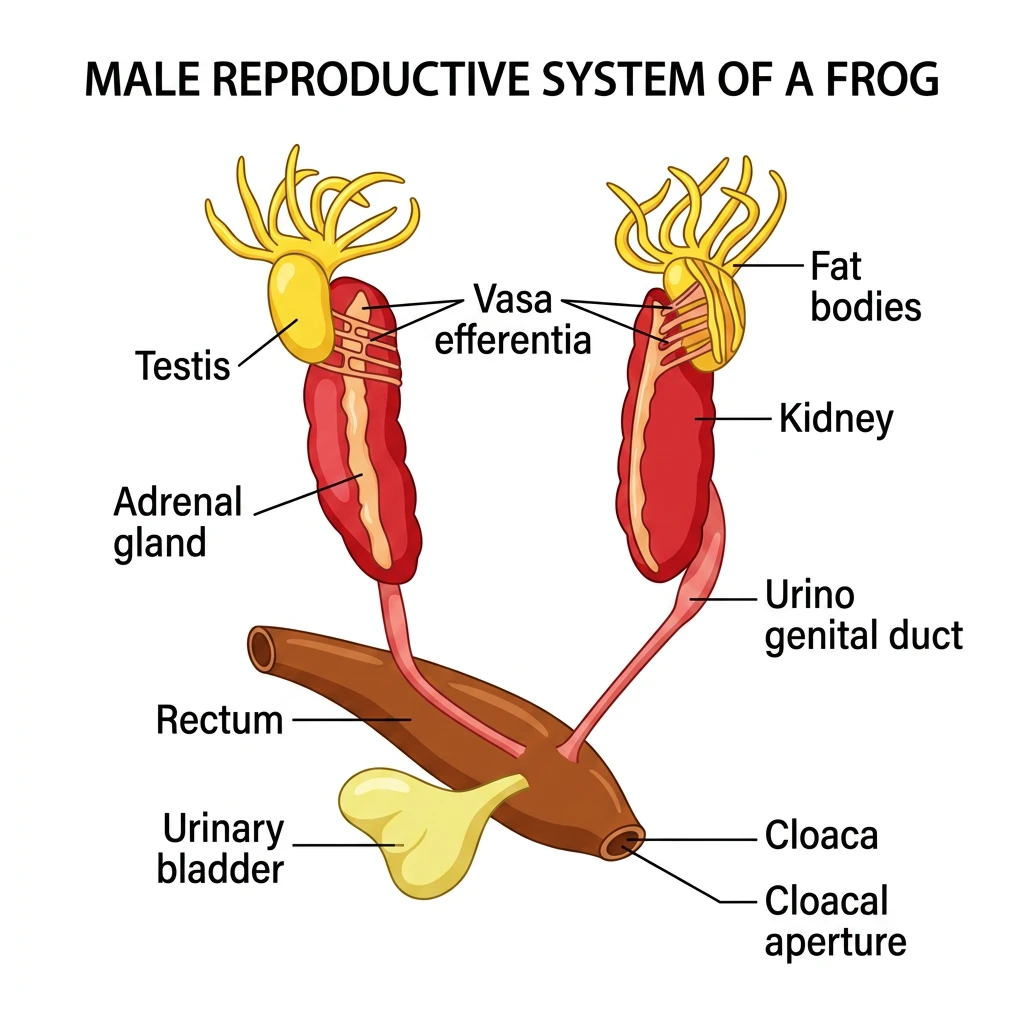
Frog Male Genital System: Details the testes, vasa efferentia, and shared urinogenital ducts.
🔬 Detailed Anatomical Description
The male reproductive organs of a frog comprise: (1) A pair of yellow ovoid testes adhered to the upper part of the kidneys; (2) 10-12 thin vasa efferentia tubes that pierce the kidneys to open into Bidder's canal; (3) Bidder's canal communicates with the urinogenital duct, which exits the kidney and runs down to open into the cloaca alongside the bilobed urinary bladder.
B FEMALE REPRODUCTIVE SYSTEM
| STRUCTURE | NCERT ANATOMICAL DESCRIPTION |
|---|---|
| Ovaries | A pair of lobed, dark grey ovaries situated near kidneys. Ovaries have no functional connection with kidneys ⭐⭐⭐ |
| Oviducts | A pair of coiled, glandular oviducts arising from abdominal cavity and opening independently into the cloaca ⭐⭐⭐ |
| Egg Laying (Oviparity) | A mature female frog can lay 2500 to 3000 black, spherical ova (eggs) at a time in water ⭐⭐⭐ |
Frog Female Reproductive System
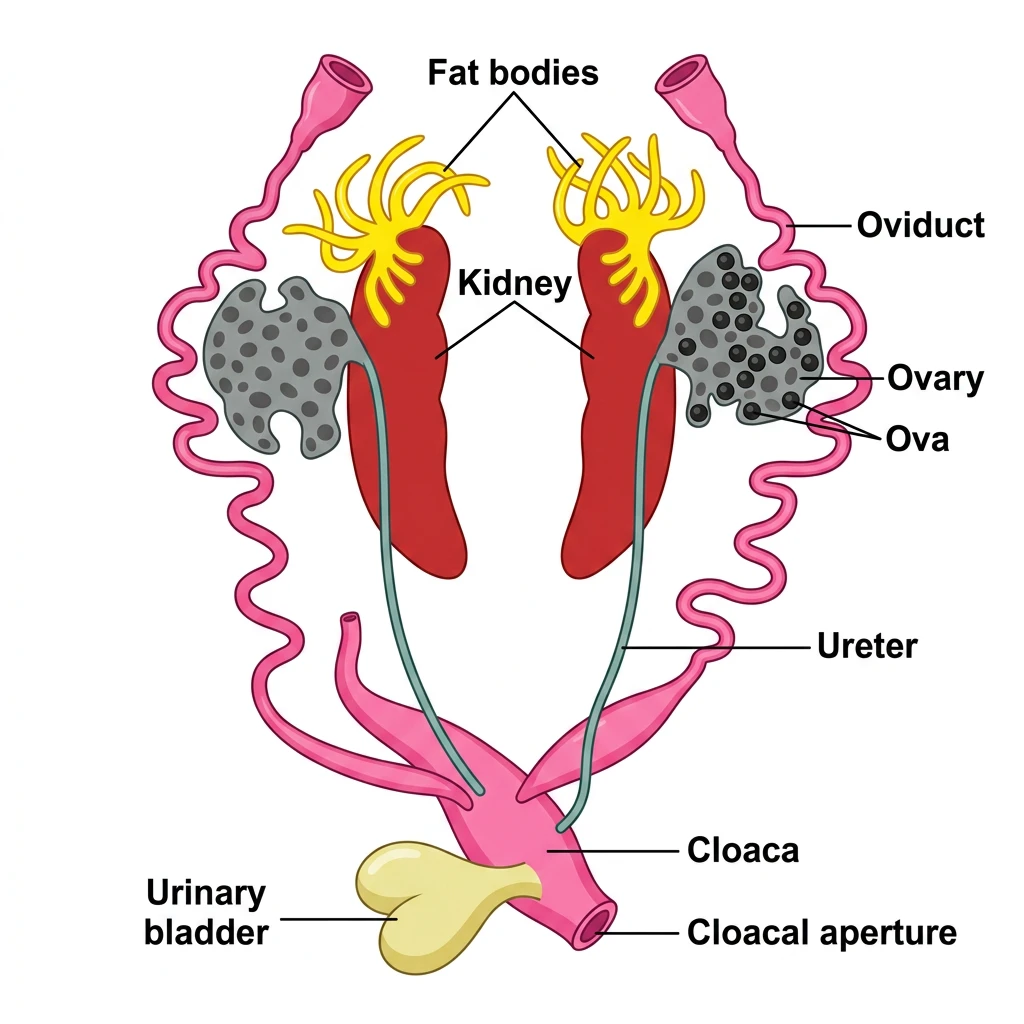
Frog Female Genital System: Shows the pair of ovaries full of ova, along with independent coiled oviducts.
🔬 Detailed Anatomical Description
The female reproductive organs of a frog include: (1) A pair of lobed, dark-grey ovaries situated near the kidneys, containing numerous mature ova (eggs) which are released into the body cavity; (2) Coiled, glandular oviducts that open independently into the cloaca; (3) Separate ureters that run along the kidneys to carry urine, opening independently into the cloaca alongside the thin-walled urinary bladder.
C FERTILISATION & DEVELOPMENT (METAMORPHOSIS)
| STAGE / EVENT | BIOLOGICAL SIGNIFICANCE |
|---|---|
| External Fertilisation | Fertilisation is external and takes place in water during breeding season (rainy season) |
| Larval Stage (Tadpole) | Development is indirect involving a free-swimming larval stage called tadpole. Tadpole is herbivorous and has gills for respiration ⭐⭐⭐ |
| Metamorphosis | Tadpole undergoes metamorphosis to form an adult frog. Metamorphosis requires thyroid hormone (thyroxine) and iodine in water ⭐⭐⭐ |
NEET 2018, 2022, 2026 (reinforced): In male frogs, vasa efferentia (10-12 in number) open into Bidder's canal inside the kidney. In female frogs, ovaries have no functional connection with kidneys and oviducts open independently into the cloaca.
XIV. FROG — RANA TIGRINA • D. ECONOMIC IMPORTANCE & ECOLOGICAL ROLE
Economic & Ecological Importance of Frogs: Frogs play a vital role in maintaining the ecosystem and are beneficial to humans in multiple ways.
| ROLE / BENEFIT | NCERT EXPLANATION |
|---|---|
| Beneficial to Mankind (Pest Control) | Frogs eat insects and pests, thus protecting agricultural crops from insect infestation ⭐⭐⭐ |
| Ecological Balance (Food Web) | Frogs maintain ecological balance as they serve as an important link in both aquatic and terrestrial food chains / food webs (acting as both predator of insects and prey for snakes/birds) ⭐⭐⭐ |
| Edible Source | In some countries, the muscular hind legs of frogs are used as food by humans |
XV. RAPID REVISION & COMMON EXAM TRAPS
⚡ RAPID REVISION — CONSOLIDATED NEET 2026 / RE-NEET 2026 EXAM TRAPS
MASTER COMPARISON TABLES — HIGH-YIELD NEET UG BIOLOGY
1. Male Frog vs Female Frog ⭐⭐⭐
| CHARACTERISTIC | MALE FROG | FEMALE FROG |
|---|---|---|
| Vocal Sacs | Present (under neck) | Absent |
| Copulatory Pad | Present on 1st digit of forelimb | Absent |
| Ureters / Ducts | Act as Urinogenital Ducts (carry both urine and sperms) | Ureters and Oviducts open independently into cloaca |
| Kidney Connection | Vasa efferentia (10-12) enter kidney and open into Bidder's canal | Ovaries have NO functional connection with kidneys |
2. Aquatic vs Terrestrial Respiration in Frog ⭐⭐⭐
| ENVIRONMENT / STATE | ACTIVE RESPIRATORY ORGANS | MECHANISM |
|---|---|---|
| In Water | Skin exclusively (Cutaneous respiration) | Dissolved O₂ exchanges with CO₂ by diffusion through moist skin |
| On Land | Skin + Buccal Cavity + Lungs | Cutaneous, Buccal, and Pulmonary respiration all operate |
| Aestivation / Hibernation | Skin exclusively (Cutaneous respiration) | Sole respiratory organ during seasonal dormancy in burrows |
3. Frog vs Human Anatomy Comparison ⭐⭐⭐
| FEATURE | FROG (RANA TIGRINA) | HUMAN (HOMO SAPIENS) |
|---|---|---|
| Cranial Nerves | 10 pairs | 12 pairs |
| Red Blood Cells (RBCs) | Nucleated, oval, biconvex | Enucleated (anucleate), circular, biconcave |
| Heart Chambers | 3-chambered (2 Atria, 1 Ventricle) | 4-chambered (2 Atria, 2 Ventricles) |
| Renal Portal System | Well-developed | Absent / Vestigial |
| Nitrogenous Waste | Ureotelic (excretes urea) | Ureotelic (excretes urea) |